Integumentary and Alimentary System. The Adult Salamander
A review of the amphibian integument was recently published (Heatwole & Barthalmus, 1994).
The Adult Caecilian. Caecilians demonstrate the typical amphibian integument that consists of very few cell layers. As a rule the stratum corneum consists of a single layer of keratinized cells. The basal epidermal cell layer is often less than eight cells thick and is underlayed by a basement membrane. The dermis lies immediately beneath this and contains capillaries, nerves, and smooth muscle throughout its two layers.
The dermis consists of an outer spongy layer and inner compact layer. Chromatophores and glands are present within the spongy layer, while the compact layer possesses collagen fibers that intimately adhere it to the underlying muscles and bones. Unlike anurans and salamanders, some caecilians possess tiny dermal scales.
Many of the glandular secretions of caecilians can prove irritating if introduced into a human's ocular membranes (O'Reilly et aI., 1995). Annuli serve to increase the surface area of the caecilian and may facilitate gas and water exchange. Shedding of the stratum corneum occurs on a regular basis. Many caecilians eat their shed skin (Weldon et aI., 1993).
The Adult Salamander. Salamanders have the typical amphibian integument, but the stratum corneum of some aquatic species is not keratinized. The dermis is firmly attached to the underlying muscles and bones.
The glandular secretions of some salamanders possess toxic compounds. Modifications such as costal grooves, granular skin, and skin folds increase the surface area of the integument and may facilitate gas and water exchange. Shedding of the stratum corneum occurs on a regular basis. Many salamanders eat their shed skin (Weldon et aI., 1993).
The Adult Anuran. Most anurans have the typical amphibian integument, but modifications of the typical plan are numerous. The skin is not as tightly adhered to the underlying structures as it is in caecilians and salamanders, and this potential subcutaneous space can fill with fluid. Thus anurans may appear edematous, a condition not described for caecilians or salamanders. Subcutaneous fluid accumulation in the anuran can be normal, functioning as a reserve of water, or it may be the result of a pathologic process. Anurans generally have a greater range of color changing ability than other amphibians.
The skin over the skull of certain anurans (e.g., bufonids) is co-ossified with the underlying dermal hones, while other frogs (e.g., giant monkey frog, Phyllomedusa hieolor) have small bones within the dermis. Similar to other amphibians, shedding of the stratum corneum occurs on a regular basis and many anurans are keratophagous (Weldon et ai., 1993).
Alimentary System.All adult amphibians are primarily carnivorous and have a relatively short and simple gastrointestinal tract. Comprehensive descriptions and reviews of the digestive tract have been published (Olsen, 1977;
Reeder, 1964).
Some mastication occurs in the oral cavity, but prey is usually swallowed whole. The oral cavity is separated from the esophagus by a strong sphincter, and the esophagus is separated from the stomach by a sphincter. Cilia lines the esophagus to transport ingesta and secreted material to the stomach. Mucous and some digestive enzymes (e.g., pepsinogen) are secreted by glandular cells lining the esophagus. The stomach is separated from the intestine by a pyloric sphincter, and generally lies to the left of midline within the coelom.
The sections of the intestine are not as grossly obvious as in other vertebrates. Gastric emptying is controlled by the duodenum. The liver and gall bladder are intimately connected, and a pancreas is present. The liver has a minimal role in processing nitrogen for excretion in aquatic amphibians, as ammonia is freely diffused into the surrounding environment through the skin and via excretion via the kidneys. In terrestrial amphibians, the liver converts ammonia to the less toxic water-soluble nitrogenous compound urea, and in a few species urea is converted to uric acid as a further method of water conservation. (See Section 4.2, Water Homeostasis.)
The pancreas is found in the hepato-gastric ligament between the stomach and anterior intestine. Bile and pancreatic enzymes enter the intestine through ducts that empty into the anterior part of the small intestine (duodenum). The small intestine is the site of enzymatic digestion and carbohydrate, fat, and protein absorption. The large intestine is the site of water and salt absorption and mucous secretion to aid in the passage of fecal boluses. Fecal matter is voided through the cloaca. Amphibian feces will contain undigested parts of the ingesta, including chitin, keratin, cellulose, and the bones that were not decalcified by the acid secretions of the stomach.
The Adult Caecilian. The arrangement and distribution of teeth are diagnostic characters in the identification of many caecilians (Taylor, 1968). Multiple rows of teeth are a common feature of many caecilians. Multiple pancreatic ducts are present, while there is no valve separating the small intestine from the large intestine.
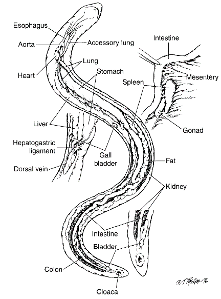
Figure 3.3. General anatomy of a caecilian. Inset view of cloaca and internal anatomy. (Tim Phelps)
The Adult Salamander. Certain oral glands are lacking in some species of aquatic salamanders (e.g., amphiumas, Amphiuma spp., sirens, Siren spp.). Pepsinogen secreting cells are absent in some sala- mandrids (Salamandra spp.). Pancreatic ducts range in number from 2-47 depending on the species. There is no valve separating the small intestine from the large intestine.
The Adult Anuran. Teeth are absent in some anurans. Pipid frogs lack tongues and certain oral glands, features that are unnecessary for their mode of aquatic suction feeding. Pepsinogen secreting cells are absent in some pipids. The liver is bilobate. Some anurans can evert their stomach and use their hands to wipe ingesta from the mucosal surface, an apparent adaptation allowing removal of indigestible or toxic substances. There is a single pancreatic duct, but the pancreatic and bile ducts may merge prior to entrance in the intestine in ranids. There is no valve separating the small intestine from the large intestine in many species of primitive anurans.
Date added: 2022-12-11; views: 1245;