Development and Structure of the Adrenal Glands
A. Adrenal Cortex. The adrenal cortex is derived from the mesoderm and can be identified in the human fetus by the age of 6 wk near the cephalic end of the mesonephros. By 2 mo, a capsule of connective tissue surrounds each gland. In the fetus, the gland is composed of an inner fetal zone and an outer definitive zone, the latter of which is similar to the adult cortex.
The fetal adrenal rapidly increases in size, largely due to growth of the fetal zone, and by midgestation is larger than the fetal kidney. In comparison to total body mass, the adrenal gland is disproportionately larger in the fetus than in the adult. At birth, the fetal zone, which produces mostly dehydroepiandrosterone sulfate (DHAS), involutes, and within days, circulating concentrations of DHAS fall. Within several months, the fetal zone is not detectable. Why involution occurs is not clear. Although the fetal adrenal is responsive to adrenocorticotropin (ACTH; see below), involution occurs in the presence of ACTH.
The location and surrounding structures of the human adrenal glands are shown in Fig. 1. The weight range of each adrenal gland is approximately 3.5-5 g, and the cortex comprises 90% of the gland volume. Chronically increased ACTH secretion causes adrenal weight to increase. Occasionally, accessory adrenal tissue is found in the connective tissue near the main glands.

FIGURE 1. Location and surrounding structures of the human adrenal glands. [Source: Gaudin, A. J., and Jones, К. C. (1989). “Human Anatomy and Physiology.” p. 423. Harcourt Brace Jovanovich, San Diego. Reproduced with permission.]
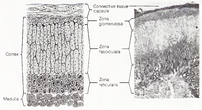
By light microscopy, three zones can be identified in the cortex (Fig. 2). The outer zone, the zona glomerulosa, is relatively thin and contains cells that secrete aldosterone. The middle zone, the zona fasciculata, is usually the thickest layer of the adrenal cortex and has a columnar structure. Its cells are relatively clear because they are large and have a high lipid content.

The inner zone, the zona reticularis, surrounds the medulla. Its cells are relatively dark staining and compact in appearance, and they often contain lipofuscin pigment granules. Both the zona fasciculata and the zona reticularis produce cortisol and androgens. Chronically increased ACTH concentrations result in lipid depletion from the zona fasciculata and an increase in the width of the zona reticularis.
B. Adrenal Medulla.In the fetus, the sympathetic nervous system arises from cells (sympathogonia) of the neural crest. By week 5 of gestation, these cells migrate from the primitive spinal ganglia in the thoracic region to form the sympathetic chain. By week 6, large groups of neural cells migrate along the central vein into the adrenal gland, and thereby form the primitive adrenal medulla. Storage granules, which stain brown with chromic acid due to oxidation of catecholamines to melanin, are found by week 12.
The storage granules give adrenomedullary cells the name chromaffin or pheochrome cells. These cells also are found on both sides of the aorta and comprise the paraganglia. The largest collection of these cells is found near the inferior mesenteric artery, where they fuse to form a fetal structure termed the Organ of Zuckerkandl, which undergoes involution within the first year of life.
The remainder of the chromaffin cells in the paraganglia and adrenal medulla persist. In the adrenal medulla, the cells are arranged in an irregular network with a rich blood supply and are in contact with sympathetic ganglia. The cells of the adrenal medulla are innervated by preganglionic fibers of the sympathetic nervous system. The blood supply of the adrenal glands enters through the cortex and drains into the medulla, except for some vessels that supply the medulla directly.
Date added: 2023-05-09; views: 823;
