Vacuum Extraction of the Fetus
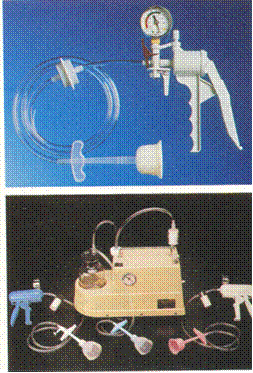
Vacuum extraction is a type of operative delivery with a help of apparatus consisting of a vacuum pump and a set of cups which are applied to the scalp of the fetal head (Fig. 234).
The operative principle consists in creating a negative pressure in the space between the fetal head and the steel cup by which the head is firmly held. Traction is applied to the cup to accomplish delivery.

Fig. 234. Two examples of a soft-cup vacuum extractor system
A vacuum extractor is used in cases where parturition should be accelerated, while a cesarean section is contraindicated. The indications for vacuum delivery are:
- persistent weakness of expulsive pains,
- progressive intrauterine asphyxia.
A vacuum extraction of the fetus cannot be used for substitution of expulsive pains (in patients with severe diseases, which require removal of abdominal contractions).
Requisite conditions: a fully dilated cervix, ruptured fetal membranes, absence of cephalopelvic disproportion, occiput presentation. It cannot be used at any deflexed presentations of the fetal head.
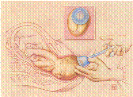
The suction cup is applied to the head away from the fontanelles but near the leading point. Vacuum pressure up to 0.7–0.8 kg/cm is reached, and traction is applied with one hand on the vacuum while the other hand maintains fetal flexion and supports the vacuum cup. Traction should be applied only during contractions. The direction of any traction depends on the head position (the same as in forceps delivery described above) (Fig. 235).
The vacuum pressure can be reduced between contractions and should not be maintained longer than 30 minutes.

Fig. 235. Illustration showing a crowning fetal head with a vacuum extractor cup attached
Destructive Operations. The destructive operations are performed to diminish the bulk of the fetus so as to facilitate easy delivery through the birth canal. There are four types of operation:
- craniotomy
- evisceration
- decapitation
- cleidotomy
Craniotomy. It is an operation to make a perforation on the fetal head, to evacuate the contents followed by extraction of the fetus.
Indications: cephalopelvic disproportions with dead fetus, hydrocephalus (sometimes other congenital anomalies) even in a living fetus, a severe general condition of parturient, the necessity of immediate termination of labor.
Requisite conditions: a fully dilated cervix, a dead fetus, the true conjugate over 7 cm, ruptured fetal membranes.
Anesthesia for relaxation of abdominal muscles and analgesia are indicated.
Contraindications: the operation should not be made when the pelvis is severely contracted (the true conjugate is 7 cm and less). In such condition the baby can be delivered only by cesarean section.
The operation consists of the following steps:
- perforation of the fetal head,
- excerebration (destruction and removal of the brain),
- cranioclasis (removal of the fetal head).
The necessary tools are a Fenomenov perforator or a Blot perforator, wide vaginal specula, elevator. Museux forceps, bullet forceps, spoon for destroying the brain or Budin’s cannula, cranioclast and scissors for cutting clavicles.
Technique. The head is steaded by assistant from the outside. Under the cover of the whole hand the perforator is introduced and applied to the most accessible portion of the head, care being taken to avoid its slipping off. It is also possible to expose the head with specula and perform the operation under direct vision. When the fetal head is exposed, a perforator should be stuck into the skull, the opening (hole) should be carefully dilated, a spoon for destroying the brain should be introduced into the cranial cavity and the brain matter is broken up thoroughly, taking special care of tearing the tentorium and destroying the medulla. Then the skull is washed out wit sterilized water. The third step of the operation consists in grasping and removing of the empty skull.
The internal blade of the cranioclast is introduced through the opening in the cranial vault and bored into the base of the skull until it is solidly fixed there, with its convexity directed towards the face. After that the outer blade is laid over the face. But after the two blades are locked and before they are tightened the physician should be assured that no maternal tissue is caught in the grasp of the jaws. Extraction is done with the same care and delicacy as a forceps operation; a proper mechanism of labor should be followed.
Decapitation. It is a destructive operation whereby the fetal head is severed from the trunk and the delivery is completed with the extraction of the trunk and that of the decapitated head per vagina.
Indication is neglected transverse presentation.
The requisite conditions are the same as in craniotomy.
Instruments: decapitation hook, blunt long-handled scissors, specula, bullet forceps for inspection of the cervix, and tool for repair of the cervical or perineal lacerations, if any.
Cleidotomy. It is a surgical division of the clavicle to facilitate passage of the shoulders through the birth canal. Both clavicles are incised, if necessary. The necessity of cleidotomy may arise after decapitation or in difficult labor of a giant fetus. The midpoint of the clavicle is located by the index and middle fingers of the guiding hand along which long-handled blunt scissors are directed to the clavicle which is incised. After the anterior clavicle has been thus divided, the shoulder girdle is decreased by 2.5-3 cm; if both clavicles are cut, the girdle is decreased by 5 or 6 cm. Extraction must be done according to the proper mechanism of delivery.
Evisceration. Evisceration (eventration) is the incision of the abdominal wall or chest of the fetus and removal of its internal organs.
Indication: neglected transverse presentation when the neck of the fetus is inaccessible for decapitation.
The requisite conditions are the same. The abdominal wall of the fetus is incised by scissors and the fetus is thus disemboweled. The proper mechanism of delivery should be followed during exraction.
Spondylotomy. It is the destruction of the spinal column. After removal of the internal organs, the spine is divided by scissors or fractured by a decapitation hook. The soft tissues are then dissected and the upper part of the fetus is extracted followed by extraction of the podalic end.
Date added: 2022-12-25; views: 789;
