Technique of the Lower Segment Operation
1. The abdominal incision should be of sufficient length to allow for delivery and may be vertical or transverse.
a. The vertical incisions are fast and can be extended above the umbilicus if more room is needed. Less dead space is present in the wound, which decreases the risk of infection. The resulting wound, however, is weaker than that from a transverse incision. The skin and subcutaneous tissue are dissected sharply down to the fascia. The fascia can be incised vertically with a knife, or a window can be created and then the incision is extended with Mayo scissors. The rectus and pyramidalis muscles are then separated at the midline, which exposes the peritoneum. The peritoneum can be entered bluntly or tented between two instruments and entered sharply, after transillumination demonstrates no underlying bowel or omentum. The peritoneal incision is extended superiorly and inferiorly, with care taken to avoid the bladder and bowel. A parietal peritoneum may be dissected in transverse direction.
b. A sufficient transverse or Pfannenstiel incision is made approximately 2 finger breadths above the pubic symphysis. The tissue is divided sharply down to the fascia, which is transversely incised in a curvilinear fashion, either with the scalpel or with scissors. The superior and then the inferior edge of the fascia is grasped and elevated, and the fascia is either bluntly or sharply separated from the underlying rectus muscles. Dissection is continued superiorly to the level of the umbilicus and inferiorly to the pubic symphysis. The peritoneum is entered in the manner described earlier. A parietal peritoneum may be dissected in a transverse or longitudional direction.
2. Bladder flap. The uterovesical fold is grasped, elevated, and sharply incised above the upper border of the bladder in the midline (about 1.5 cm above the apex of the bladder). Then the fold should be opened in each direction to undermine the serosa before sharply incising it. The bladder and lower portion of the peritoneum are then bluntly dissected off the lower uterine segment, and the lower segment is exposed. Then a bladder blade may be replaced between the bladder and the lower uterine segment (Fig. 220 C, D).
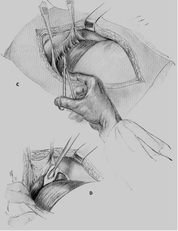
Fig. 220. Cesarean section. The bladder is exposed (C) and lower uterine segment is exposed (D)
3. The transverse incision is used most commonly. A curvilinear incision is made transversely in the lower uterine segment at least 1–2 cm above the upper margin of the bladder (Fig.220 E). The uterine cavity is entered carefully in the midline, with care taken to avoid injury to the fetus. The length of this insicion is 2 cm only. Then this incision should be extended bilaterally with two fingers to the sides and upward (Fig 220 F), with care taken to avoid the uterine vessels laterally. This type of incision is associated with less blood loss, fewer extensions into the bladder, decreased time of repair, and lower risk of rupture with subsequent pregnancies than other types of incisions. Disadvantages are the limitation in length and greater risk of extension into the uterine vessels.
The advantage of the low vertical incision is that it can be extended if more room is needed; in so doing, however, the upper segment of the uterus may be entered. Such an occurrence should be recorded in the operative notes, and the patient should be informed and counseled that vaginal birth trial is contraindicated thenceforth, as the risk of uterine rupture is as high as 9%. Low vertical incisions are associated with extensions into the active segment more frequently than transverse incisions. In addition, to avoid injury, the bladder must be dissected further for low vertical incisions than for transverse incisions.
The classical type of incision extends from 1 to 2 cm above the bladder vertically up into the upper segment of the uterus. Classical incisions are associated with more bleeding, longer repair time, greater risk of uterine rupture with subsequent pregnancy (4–9%), and greater incidence of adhesion of bowel or omentum. In cases of fetal prematurity, lower uterine segment fibroids, malpresentations, or fetal anomalies, however, it may be necessary to make this type of incision to provide adequate room for delivery.
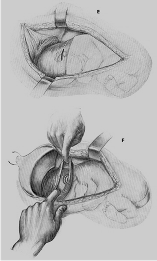
Fig. 220. Cesarean section: E - dissection of the uterine wall; F - the incision is extended laterally and upward; .M, N –the head is elevated through the incision
The head is elevated through the incision. The remainder of the fetus is delivered using gentle traction on the head as well as fundal pressure. The child is extracted either by the leg, if the breech presents or by grasping the head with one hand (Fig. 220 M, N). Shoulders and trunk should be removed after it (Fig. 220 O, P). If it is indicated the infant may be delivered with the forceps, or vacuum extraction (more rarely). (Fig. 220 K, L). The infant's nose and mouth are suctioned, the umbilical cord clamped and cut between two forceps (Fig. 221), and the infant is delivered to the resuscitation team. The placenta is removed manually (Fig. 222).
After the delivery of placenta, oxytocin is administered. The uterus may be removed through the abdominal incision or left in its anatomic position. The incision is inspected for extensions, and the angles and points of bleeding are clamped with fenestrated clamps. The uterine cavity is wiped with a laparotomy pad to remove the retained membranes or placental fragments; some surgeons prefer to perform the curettage of the uterine cavity to prevent remaining of placental and membrane tissue in the uterus.
Repair begins laterally to the angle of incision, with care taken to avoid the uterine vessels. A running or running locking stitch is placed. The entire myometrium should be included. The second imbricated stitch, either horizontal or vertical, may then be placed if hemostasis is not obtained with the initial suture. The edges of the divided uterovesical fold of the peritoneum are then united with a continuous catgut suture (Fig.223).
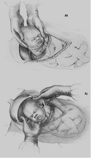
Fig. 220. Cesarean section: M,N –the head is elevated through the incision
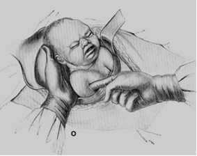
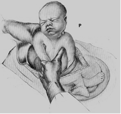
Fig. 220. Cesarean section: O - removing of shoulders; P - removing of trank
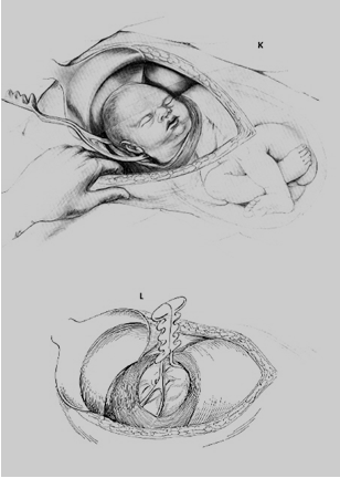
Fig. 220. Cesarean section: removing of the head by forceps
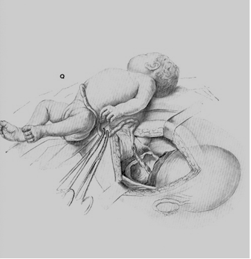
Fig. 221. Cutting of the umbilical cord between two clamps
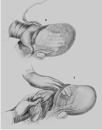
Fig. 222. Removing of the placenta
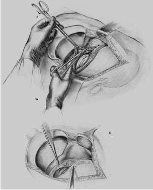
Fig. 223. Repairing of the uterine wound
The incision is inspected, and further areas of bleeding may be controlled with figure-of-eight sutures or electrocautery.
In classical cesarean sections, two or three layers of sutures may be required to close the myometrium. The serosa should then be closed with an inverting baseball stitch to decrease formation of adhesions of bowel and omentum to the uterine incision.
Abdominal closure. The tubes and ovaries are inspected. The posterior cul-de-sac and gutters are cleaned of blood and debris. The uterus is returned to the anatomic position in the abdominal cavity and the incision reinspected to assure hemostasis with the tension off the vessels. The fascia is then closed with running delayed-absorption sutures. The subcutaneous tissue is inspected for hemostasis, and dead space may be closed with interrupted absorbable sutures. The skin is closed with subcuticular stitches or staples.
Date added: 2022-12-25; views: 757;
