Supplemental Examinations Using Conventional and Modern Imaging Techniques
Panoramic radiography offers the dentist a realm of diagnostic possibilities that will lead to additional knowledge and a deeper understanding of anatomic relationships. This will provide new therapeutic insight, and will lead the dental practitioner to an awareness of the necessity to become expert not only with panoramic radiography and its diagnostic possibilities but also with supplementary conventional and more contemporary methods of imaging.
For many and varied reasons it may seem impossible for the dentist to possess and routinely use all of the possibilities afforded by modern techniques for examination in his or her practice; it is nevertheless necessary that practitioners be aware of the more important examination techniques and the diagnostic possibilities that they offer so that the dentist can advise and refer patients appropriately.
Today it is possible to take advantage of numerous diagnostic possibilities offered even by panoramic radiography, because modern electronics in the newer instruments permit the creation of many targeted projections in addition to the normal panoramic film. When used properly, for example as a final check after completed therapy, the radiation dose to patients at follow-up examinations can be significantly reduced. However, no new information will be gleaned from targeted projections because the direction of the central ray projections is not altered.
More interesting from a diagnostic point of view is the possible development of lateral cephalometric equipment into a dental skull instrument, because only the inclusion of the third dimension can provide any significant contribution toward perfecting dental examination technique. Most modem instruments combine the choice of various targeted projections with the possibility of spiral tomography in the third dimension. This chapter will provide additional examples of the projections described on p. 24.
There can no longer exist any doubt that today’s dentist must possess knowledge about dentogenic diseases of the maxillary sinus as well as occlusion-related lesions of the temporomandibular joint; the inclusion of these regions in the dentist’s diagnostic considerations has become mandatory. Therefore in this chapter we will present examples of the range of possibilities offered by conventional tomography as well as computed tomography (CT), insofar as they affect the dentist’s sphere of operation. In addition, the dentist must be aware of the diagnostic possibilities offered by magnetic resonance imaging (MRI) for the depiction of lesions of the disk in the temporomandibular joint, even though today we still remain far from the time when this technique may be viewed as a mandatory supplement to clinical examinations.
Conventional Skull Films. First Standard Projection: Posteroanterior Skull Projection. All skull projections, regardless of the methods by which they are produced, may be basically included in the scheme of the three standard projections:
1. posteroanterior skull projection
2. lateral skull projection
3. axial skull projection
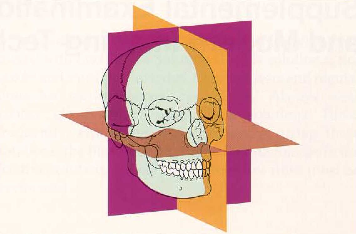
Ail of the possible skull projections originate from the three standard projections. The diagram shows the frontal plane, the median sagittal plane and the axial plane, which orients on the Frankfurt line.
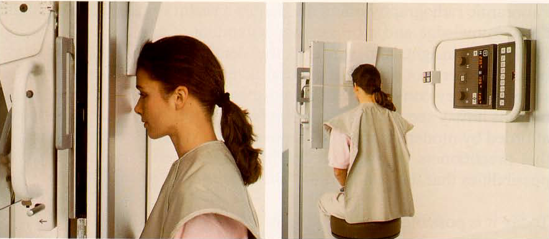
Positioning for the first standard projection, the skull overview with posteroanterior central ray projection. The petrous portions of the temporal bones are projected in the orbits.
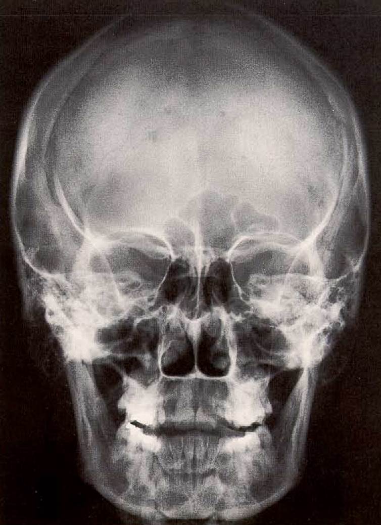
Typical “posteroanterior skull overview” radiograph. In orthodontics and in oral and maxillofacial surgery, this radiograph is used primarily for depiction of cranial asymmetries. (Radiographic anatomy of the skull radiographs may be reviewed in Pasler 1989).
Second Standard Projection: Lateral Skull Projection. Following general use in medical radiology, the second standard projection (lateral skull overview) is taken with the right side of the head against the film cassette (Fig. 309). The central ray is directed through the sella turcica. The lateral cephalometric radiograph in common use in dentistry is taken as a partial skull film with the central ray targeted throught the region of the auditory foramen. The head may be positioned with either the left or right side against the cassette.
This radiograph is used primarily in orthodontics and oral surgery. However, it may also be used in prosthodontics to check on esthetic and functional characteristics of the anterior region, and the head profile. As a lateral view of the anterior tooth region of the facial skeleton, it serves as an ideal supplement to panoramic radiography to depict the anterior tooth region. Thus this radiograph can be important for determining the spatial characteristics of cysts, tumors and impacted teeth in the paramedian region.
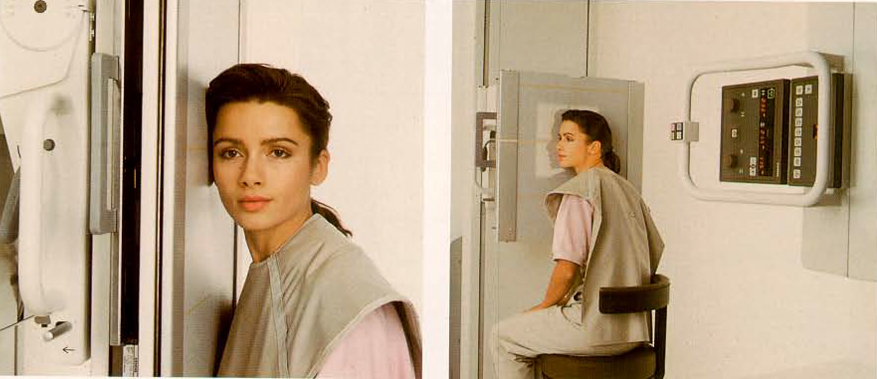
309. Positioning for the lateral skull projection. Normal position in radiology. The usual focus-film distance in radiology for skull projection is 1 m, but lor dental cephalometric projections it is 1.5 m.
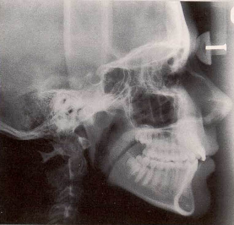
Typical cephalometric radiograph. In this case the film was taken with the left side against the cassette, with the use of a special soft tissue filter made of aluminum. The focusfilm distance of 1.5-2 m and the careful fixation of the skull in the cephalostat permits one to mark measuring points directly on the film and allows subsequent evaluation using cephalometric analysis.
Date added: 2022-12-17; views: 878;