Localization Using Various Methods
Usually the position of impacted teeth and retained root tips, but also those of foreign bodies and fracture lines, must be determined before or during appropriate therapy. During endodontic therapy, “localization radiographs” are often necessary, especially for interim examination and for follow-up. Frequently the so-called eccentric projection is appropriate, wherein the central ray is projected toward the mesial (mesial eccentric radiograph) or toward the distal (distal eccentric radiograph).
Such films are supplemental to the standard orthoradial periapical film. It will not be possible here to present examples of all possible indications for determination of the position of structures in radiographs. The principle will be presented using several typical cases of retained and impacted teeth.
In general, the following guidelines should be observed:
1. An object whose position must be localized precisely should be radiographed together with either an obvious reference object or with a characteristic anatomic structure. The basis for localization technique is a change in positional reference that results from altering the central ray projection.
2. The object-film distance of the two objects provides clues:
- Objects closer to the film appear sharper and of actual size.
- Objects distant from the film appear blurred and enlarged.
3. If possible, the object in question should be radiographed using at least two different central ray projections that are perpendicular to each other. This is usually only possible through the use of whole skull radiographs.
4. If the object in question and its reference object cannot be examined as described in point 3 above due to technical reasons, two periapical films may be employed. The first is taken as a standard orthoradial projection, while the second employs a horizontal or vertical change in the central ray projection (Clark’s technique). The apparent “movement” of the object in these two radiographs will provide clues as to its exact location.
5. Panoramic radiography extends these well-known possibilities. Objects that lie in front of the image layer appear blurred and reduced in size, while those that are behind the film appear out of focus and enlarged.
If the distance between the object in question and the reference object is too small, all methods for radiographic localization will fail, thus giving an indirect indication that the two objects are in fact in close proximity. Skull films are indispensable in many cases because they permit not only definitive changes of the central ray projection but also provide a clear overview.
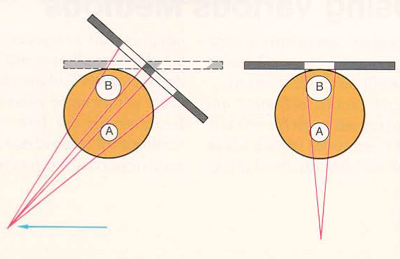
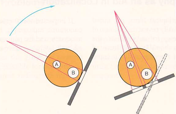
Localization using horizontal shift of the central ray projection. Schematic depiction of the orthoradial (right) and the distal eccentric (left) projections to localize impacted tooth 13 (B). The film packet need not be positioned identically for both projections. Only the proper central ray projection and the complete depiction of the crown of the tooth in question are important to compare its position to the reference object (A).
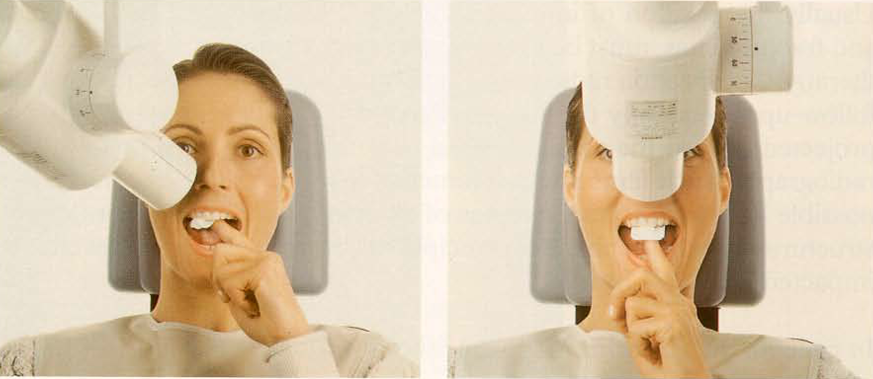
Set-up for horizontal shift of the central ray for localization of tooth 13. Note the differing film position and the distal eccentric shift of the central ray. A film holder substantially simplifies positioning of the film. This same localization technique can be used with multirooted teeth to provide radiographic evaluation of individual roots.
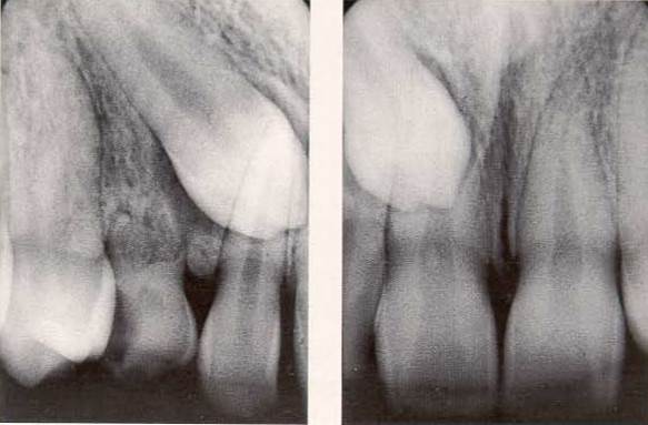
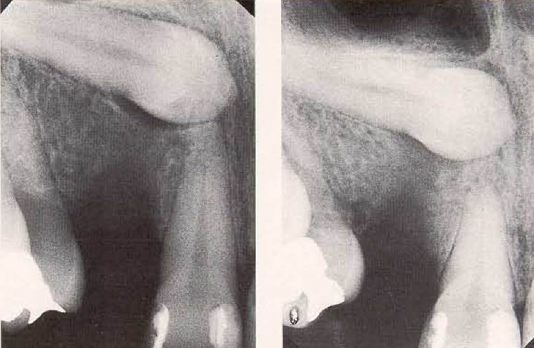
Examples of results, as viewed in radiographs. Right: Result with the initial standard orthoradial projection; left: result after movement of the X-ray tube into the distal eccentric position. A rule of thumb is that objects which move with the central ray movement are actually behind the reference object (Hotz). Thus, tooth 13 is positioned palatally.
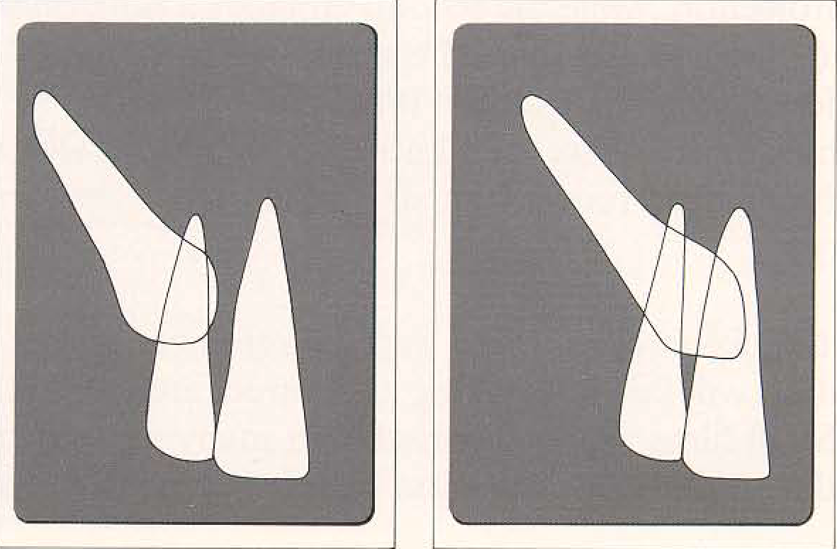
Diagram of the horizontal shift of the central ray. The crown of tooth 13 appears separate from tooth 11 (left) when the central ray is projected using the distal eccentric technique. If tooth 13 were positioned buccally (right), it would appear enlarged and would appear superimposed upon the root of tooth 11 in the radiograph.
Localization by vertical shift of the central ray. Schematic depiction of a standard orthoradial (left) and a steep projection (right) to localize an impacted tooth (B). The films need not be identically positioned for the two exposures. Most important is the proper central ray projection for depiction of the crown of tooth 13 (B) and the depiction of the root tip of 11 (A).

Set-up for the vertical shift of the central ray for localization of tooth 13. Note the vertical shift of the central ray in the right picture. This technique can be simplified by use of a film holder affixed to the end of the X-ray tube.
Result as seen in the radiograph. Left: Result with the initial standard orthoradial projection. Right: Result after vertical shift of the X-ray tube. These two films reveal that tooth 13 is positioned palatally, according of the rule of thumb that objects which appear to move with the central ray are located behind the reference object (Hotz).
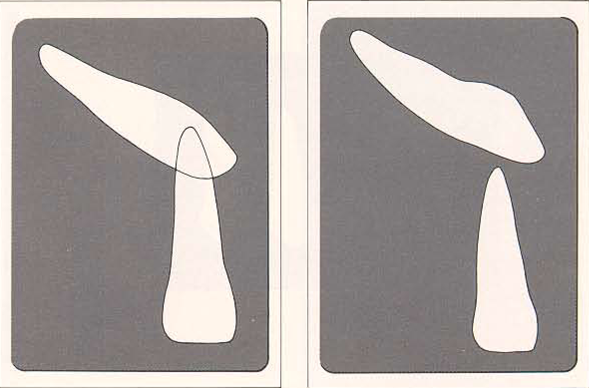
Diagram of vertical central ray shift. The central ray, which enters from above at a high angle (right) provides a radiograph that shows a separation between the crown of tooth 13 and the root tip of 11. If tooth 13 were located buccally (left), it would appear enlarged and would significantly overlap the root of tooth 11.
Date added: 2022-12-17; views: 814;