Panoramic Radiography as an Aid in Localization
In contrast to individual periapical films, the initial panoramic radiograph will usually reveal the position of impacted teeth. Nevertheless, to substantiate the diagnosis, the panoramic film should be supplemented with an occlusal radiograph or a periapical film in some cases.
If impacted teeth appear in an axial location in the panoramic radiograph, however, the greatest care is necessary, and the panoramic film must be supplemented with lateral, posteroanterior or axial skull projections.
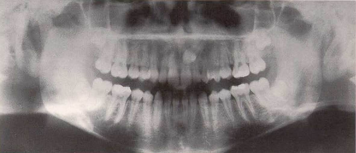
Palatal position of tooth 23, viewed in the panoramic radiograph. A comparison of the size of tooth 23 with the reference object (tooth 13), which is aligned within the dentition clearly reveals the slight enlargement of the crown and the root, indicating a greater objectfilm distance.
Palatal position of tooth 13 as seen in the panoramic radiograph. Tooth 13 is not sharply depicted and its crown appears greatly enlarged, indicating that tooth 13 is impacted palatally at great distance from tooth 11 and its root tip is located high and ventrally in the canine fossa.
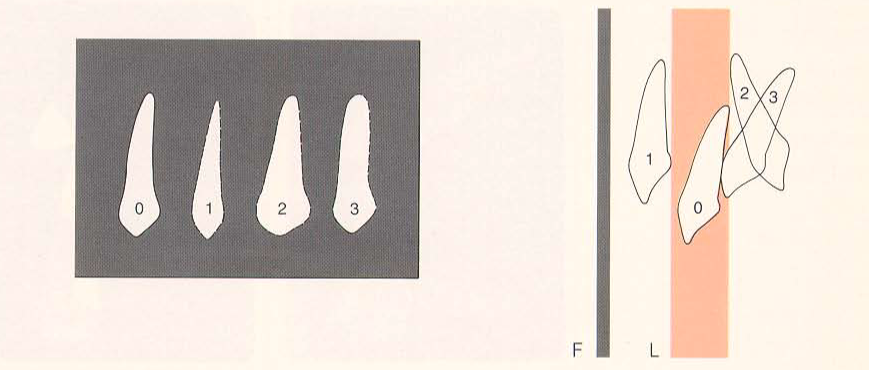
Diagram of the apparent variation of shape of impacted maxillary canines, as seen in the panoramic radiograph. The incisors or the normally developed canine (0) of the contralateral side serve as reference objects and are shown sharply and of normal size when in the image layer (L). If the impacted tooth is located buccally, it appears out of focus and reduced in size (1). If all or part of the impacted tooth is located palatally (2 or 3), it will appear blurred, and part of it will appear enlarged (distant from the film). F = plane of the film.
Buccally Impacted Mandibular Canine. Whenever panoramic radiographs must be supplemented with occlusal films for purposes of localization, one should attempt to project the group of teeth in question axially. If this is not possible for anatomic or technical reasons, the panoramic radiograph must be supplemented with appropriate periapical radiographs or skull films. Often succes is achieved only after numerous projections; such additional radiographs must be planned, however, and carried out in a logical sequence.
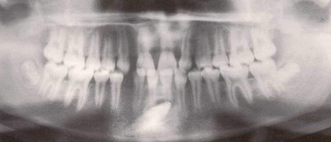
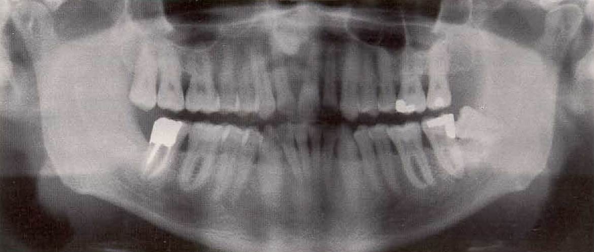
Section from a panoramic radiograph of a 13-year-old female. In addition to the congenitally missing teeth 18,12, 21 and 33, this film reveals that tooth 43 is impacted, apparently in a buccal location; it appears superimposed on the root tips of teeth 41 and 31. This projection does not allow precise localization of the impacted tooth because it appears enlarged as a result of the axial rotation and therefore simulates a lingual position (large object-film distance).
Ectopically Positioned Anterior Teeth. Whenever any individual tooth is missing (count the teeth!) or when crowns of teeth appear tipped or abnormally spaced, an absolute indication exists for a radiograph that can provide an overview, to ascertain any abnormalities of tooth development or pathological processes in the jaws as early as possible. A timely and complete radiographic examination may help to prevent later additional radiation exposure and complex therapy. In addition to panoramic radiography, in the maxilla extraoral radiographs using the occlusal film are helpful, as are skull films (e.g., lateral cephalometric radiographs).
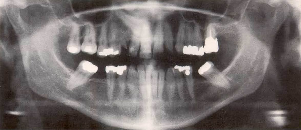
Panoramic radiograph of a 46-year-old female. This film reveals only a vague radiopacity with the approximate density of tooth substance adjacent to the anterior nasal spine. Careful examination reveals that tooth 11 is missing.
Date added: 2022-12-17; views: 841;