Apical and Periodontal Survey in Adults
The following pages present a 14-film survey for adults, using a film holder to eliminate the disadvantages associated with free-handed positioning; this technique is applicable equally for apical as well as for periodontal radiographs. In addition to the simple and practice- proven “Emmenix” film holder (Hager Co.), the preparation of apical and periodontal projections will be demonstrated with a targeting device that can provide significant enhancement of radiographic quality in comparison to free-handed positioning methods.
This is achieved via a system in which the film is held in a sterilizable and interchangeable film holder attached to an adapter ring that can be connected to the tube of almost any dental radiographic equipment. This guarantees that the film is always at a right angle to the central ray and fixed in the center of the perfectly shielded X-ray beam; in addition, this insures proper positioning of the unit and the film, as well as maximum radiation protection with each exposure.
Depicted here is the right-angle system described by the author and manufactured in Germany by Beycodent.
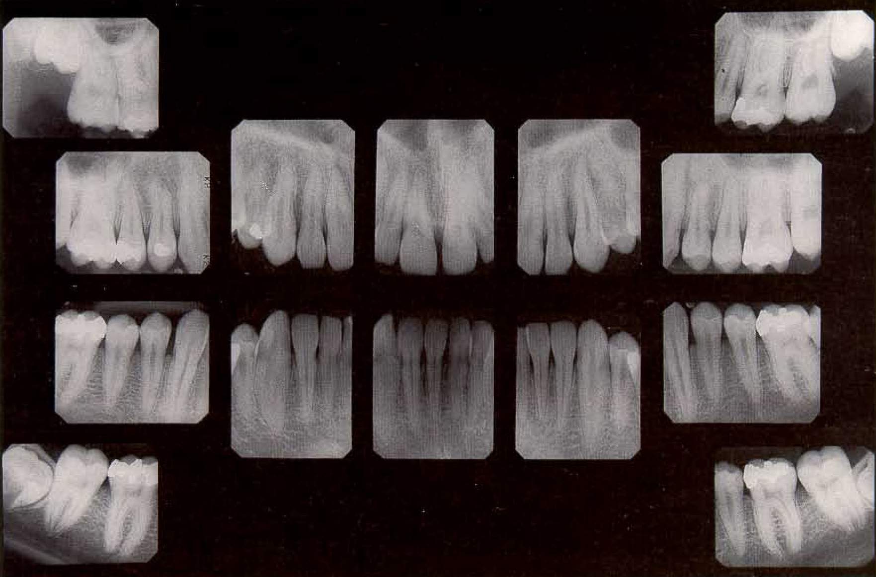
Apical 14-film survey for an adult. The central ray is targeted onto the apex; depiction of the alveolar crest is of only secondary importance. The third molars must always be located and depicted through use of special projections if no panoramic radiograph is available.
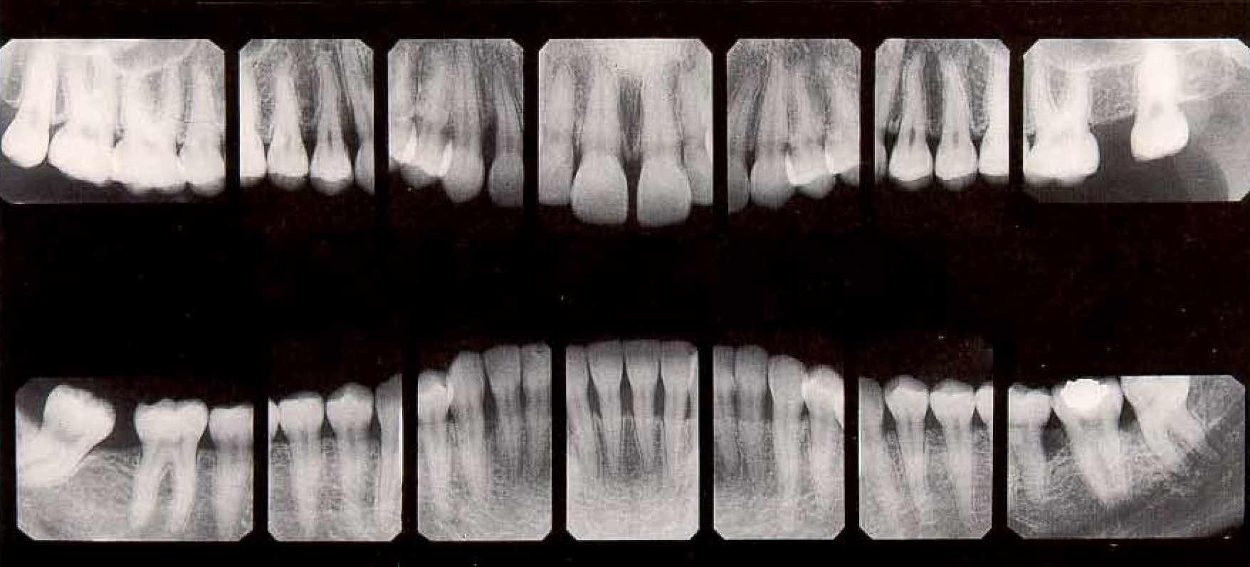
Periodontal 14-film survey for an adult. The central ray is targeted onto the alveolar crest; depiction of the root apices is of only secondary importance. The exposure data are reduced.
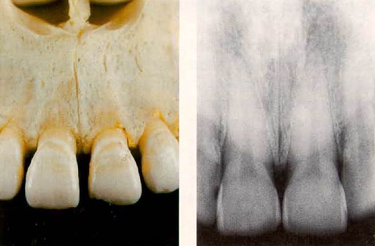
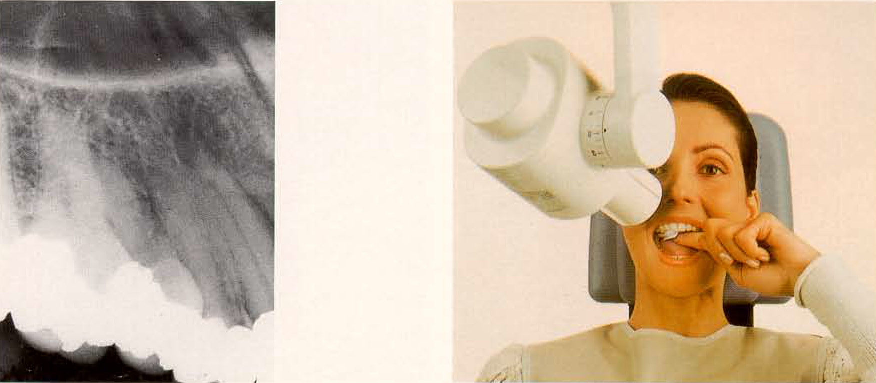
Photograph and radiograph of the region. Depending upon the indication and the age of the patient, either a 2 x 3 cm or a 3 x 4 cm film is selected and used vertically.
Film holder for the right- angle technique, as modified by Pasler. This film holder consists of a rotatable adapter ring and four interchangeable metal film holders, which serve simultaneously for targeting. Two are for the four posterior tooth regions, and two (in different sizes) are for the anterior tooth regions. The film is fixed in a right- angle configuration to the central ray, and can be rotated in the adapter ring. Radiation exposure is reduced to a minimum by incorporation of a lead collimator.
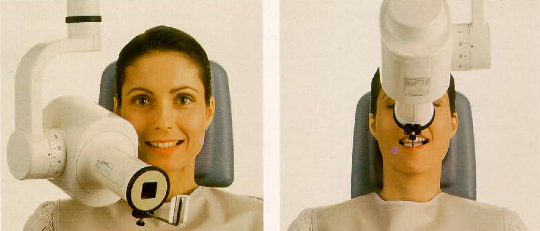
Positioning for the maxillary anterior region. Frontal view. Note the head position and the symmetrical positioning of the apparatus.
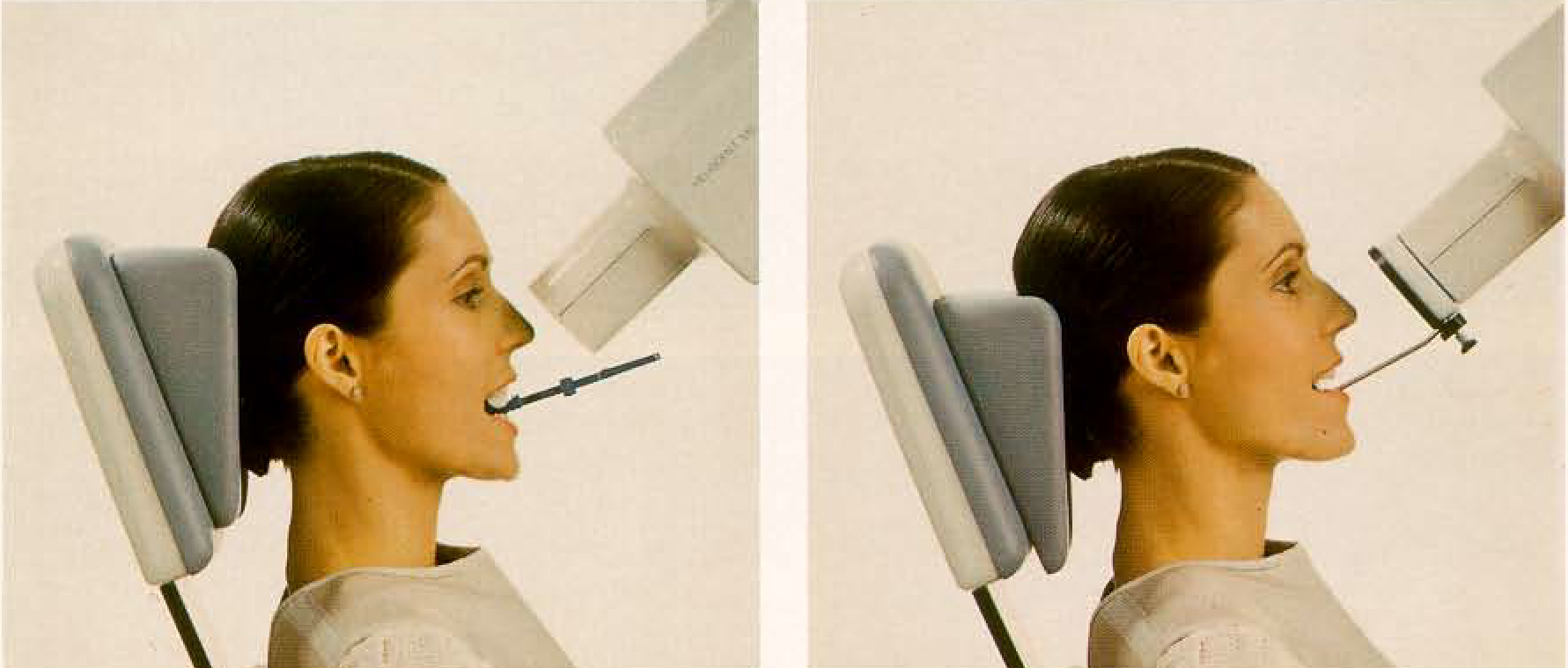
Positioning for the maxillary anterior region. Lateral view. The left picture shows the use of the Emmenixfilm holder, while the right picture shows the positioning with the film holder of Pasler. Note also the head position for radiography in the maxilla. "Snap-a-ray” is a similar model produced in the USA by Rinn Co.
The preparation of a patient for the production of a full radiographic survey or only a single radiograph is of considerable psychological importance. It is useful to insert the film initially and allow the patient to relax. Observe the patient’s reaction. Use topical anesthetics sparingly and in a targeted manner. Pay attention to proper head position: For maxillary radiographs, the bipupillary line and the occlusal plane of the maxilla should be horizontal. Select the proper horizontal and vertical angles for the projection according to the individual circumstance rather than blindly adhering to schematic tables.
Of major importance is the appropriate choice for horizontal and vertical tube angulation. In the posterior region of the jaw, the horizontal angle should be similar to an orthoradial projection to avoid the incorrect mesial eccentric positioning. At the same time, however, the vertical angle (p. 57) must be properly selected to avoid distortions of the teeth as well as summation effects with other structures. A targeting device (film holder) facilitates focusing on the critical procedures (Fig. 120).
A film holder allows placement of the film packet without having to bend it into a position dictated by anatomy. It is incorrect to press the film against the teeth and the alveolar process. In the maxilla, periapical film packets should be placed in the middle of the palate, and in the mandible toward the tongue. Therefore, in the mandible the film is not positioned anteriorly, but in the depth of the floor of the mouth. When the patient is asked to close the mouth, the musculature of the floor of the mouth relaxes and this simplifies placement of the film. Topical anesthetic may be used in the maxilla.
Date added: 2022-12-17; views: 811;