Errors in Technique that Reduce Radiograph Quality
Retaking radiographs exposes the patient to unnecessary additional radiation. Therefore extreme care should be exercised in every case to avoid errors that compromise high quality interpretable radiographic documentation. Every patient has the right to expect proper use of modem radiographic methods that guarantee minimum X-ray exposure. Inadequate knowledge or experience is an unacceptable excuse for utilizing radiographic examination as an experimental exercise.
Taking a perfect dental radiograph is not comparable to “instant photography,” rather it is demanding and requires time. Radiographs taken under constraints of time are often of poor quality. Personnel charged with taking radiographs must follow directions from their superiors and comply with all regulations concerning protection from radiation; on the other hand, the dentist must provide precise instructions for taking radiographs and must supervise their realization with responsibility and competency.
With regard to panoramic radiography, the most common errors may be traced to positioning, first of all positioning of the patient in the apparatus and secondly positioning of the patient’s head. With older units it is also possible to affix the film cassette improperly. Movement of the head or the mandible during exposure can lead to films that invite incorrect interpretation; it is for this reason that patients must be instructed before films are taken and observed during the exposure. Foreign bodies such as jewelry or a carelessly placed lead apron will lead to excessive radiation exposure if the film must be retaken; it may lead to incorrect interpretation. It is very important for perfect depiction of the maxilla that the patient press the tongue against the palate during the entire radiographic exposure; this has already been described in the section on radiographic techniques.
This tongue placement, unusual for most patients, should be practiced before positioning the patient in the apparatus. Dental prostheses, especially metal partial dentures, may be either removed or allowed to remain in the patient’s mouth, depending upon the indication, for example to clarify a temporomandibular joint disturbance or to evaluate traumatic impact. Similarly, full dentures are usually left in situ for radiographic depiction of the alveolar ridge of edentulous patients; this makes use of the filtering effect of prosthesis materials.
During the preparation of periapical radiographs, careless placement or bending of the film packet and incorrect projection of the central ray in both vertical and horizontal angles represent the most common sources of error. It is therefore highly recommended that an appropriate film holder be used to provide reproducibility and to enhance patient comfort.
Errors related to radiograph exposure time are actually less common today than is usually assumed, because of the automatic exposure devices on modern apparatus.
Before Positioning the Patient in the Apparatus.
- Take the time to explain the apparatus and the film cassette rotation to the patient
- Set the appropriate exposure data
- Have the patient remove all jewelry from ears, hair and neck
- Demonstrate the bite holder for the patient; he or she should practice biting in centric relation
- Have the patient practice proper tongue position
- Depending upon the indication, either remove the patient’s prostheses or leave them in situ
- Apply the protective lead apron to the patient
- Utilize appropriate barrier techniques
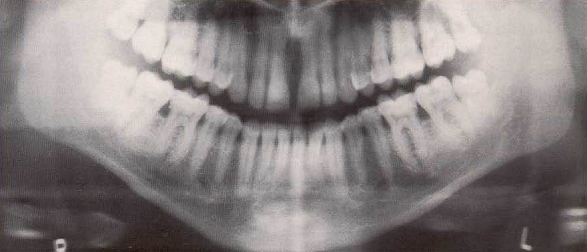
Improper positioning of the patient: Head tipped too far forward. Note the superimposition effects in the premolar regions of the maxilla. The temporomandibular joints are projected completely out of the film.

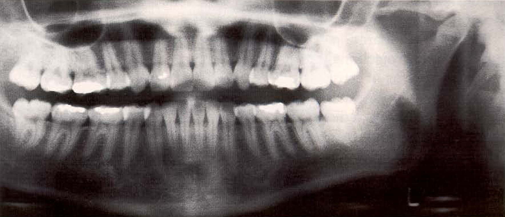
Improper positioning: Head tipped too far back. Note the superimposition on the maxillary alveolar process by structures of the floor of the nose and the palatal roof. The temporomandibular joints are projected far laterally.
Old equipment. When using older equipment, the film cassette must be placed precisely in the initial position before positioning the patient in the apparatus to preclude loss of important radiographic information. Failure to do so will result in the necessity to retake the radiograph, and therefore to additional radiation exposure for the patient.
Date added: 2022-12-17; views: 705;