Anomalies of Dental Development and the Teeth
Endogenous as well as exogenous factors can directly or indirectly elicit abnormalities of the dental lamina, which develops in close harmony with the surrounding tissues. Viewed in this way, the goal of this chapter is to summarize a large number of anomalies of the teeth that the dentist in daily practice may encounter as isolated cases or as serendipitous radiographic observations.
The list of abnormalities is long, and includes, for example, hyperodontia, hypodontia and anodontia, as well as the persistence and the inclusion of deciduous teeth, the retention of permanent tooth buds and the tooth primordia of supernumerary teeth such as the mesiodens, supernumerary premolars of the mandible and supernumerary molars of the maxilla. Such abnormalities can be considered as odontoma-related forms.
Also included are dysplasias of the tooth crowns such as dens in dente, double tooth buds and “twinning” of the tooth crowns, as well as abnormal development of the tooth roots such as concrescence and the taurodont. Concrescences of maxillary molars call to mind the complex odontoma of this region that occurs preferentially in women.
Amelogenesis imperfecta or hereditary enamel hypoplasia assumes a particular place in the category of anomalies of dental development, as do the other enamel hypoplasias with their sex-linked inheritance pattern of incomplete dominance as an ectodermal odontopathy. The radiographic examination of such lesions is of importance mainly for forensic reasons, and is of little diagnostic significance.
On the other hand, osteogenesis imperfecta is a mesodermal malformation with simple dominant inheritance. It is characterized by dentin malformation and by shortened roots and altered root form. Anomalies of this kind may be associated with corresponding developmental anomalies of the skeleton, and it may therefore be more reasonable to discuss them under the rubric “osteopathy.” The features of such lesions are usually only described in radiographic terms, because a targeted histologic examination is usually not performed in a living patient.
Numerous syndromes are accompanied by odontodysplasias. Their appearance in radiographs should therefore always lead the practitioner to a careful search for their origins. The various forms may be reviewed in the appropriate literature.
Most developmental malformations can only be correctly diagnosed by means of a complete radiographic examination of the dental structures. For definitive diagnosis in the individual case, the use of panoramic radiography as well as foil-free films for targeted or special projections is recommended.
Congenitally Missing Teeth, Retention and Inclusion. We have selected three typical examples from the myriad of cases: The relatively common congenitally missing second premolar, which often leads 1) to the persistence of the second deciduous molar, 2) the tipping of the first permanent molar and the other molars, and 3) the inclusion of deciduous molars in their original location at the deciduous occlusal plane. Note that retained deciduous molars and retained tooth buds of permanent molars must be followed up using panoramic radiography at regular intervals if surgery is not immediately indicated.
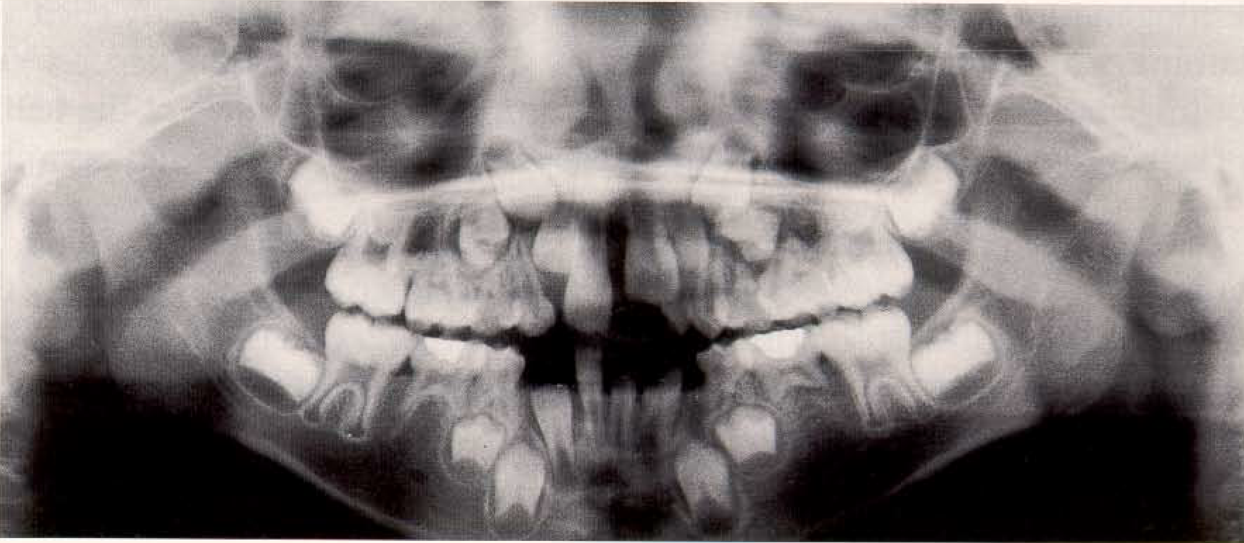
Hypodontia in a 7-year- old female. Note the complete congenital absence of all permanent tooth buds of the second premolars and the retarded eruption in the maxillary and mandibular anterior areas.
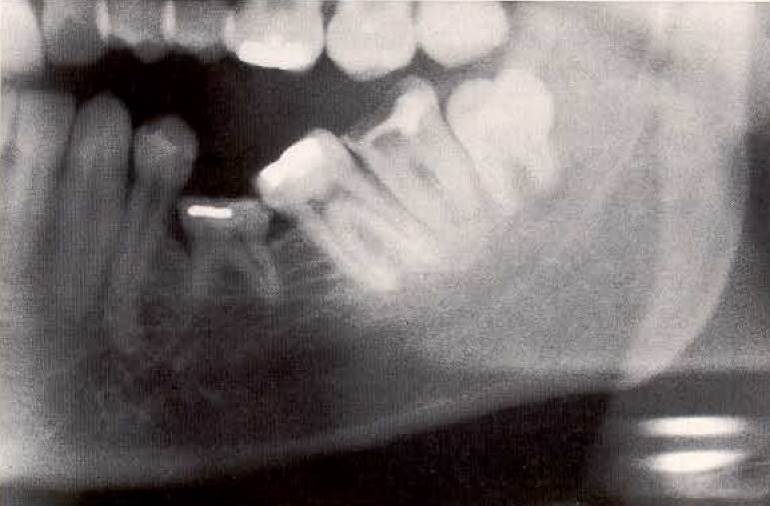
Inclusion of a persisting deciduous tooth 75. Permanent tooth 35 was congenitally absent. Note that tooth 34 as well as teeth 36, 37 and 38 have tipped toward the deciduous molar in this 23-year-old female. As a consequence of a tongue thrust habit, this patient also exhibited a lateral open bite.
Date added: 2022-12-17; views: 1047;