Rupture of the Cervix. Rupture of the Uterus
Because of effacement and taking up the cervix, which occur in the first stage of labor, the margins of cervical os are strongly thinned. Superficial lacerations of the cervical os do not frequently cause marked bleeding and usually remain unidentified. But the uterine cervix may also rupture which occurs especially often in pathological labor. The rupture is accompanied by considerable bleeding and other unfavourable consequences.
The cervix ruptures on its lateral sides, especially on the left one. The rupture may extend to the vaginal fornix, pass into it, and reach the parametrial tissues. Three degrees of the cervical rupture are distinguished: in the first degree the cervix ruptures to a length not more than 2 cm; in the second degree this exceeds 2 cm but does not reach the fornix by 1 cm; in the third degree the ruptured area reaches the fornix and passes onto it (onto the upper part of the vagina).
Blood vessels are injured in the deep rupture of the cervix to cause bleeding which is often profuse and endangers the mother’s life. The bleeding is normally external; if the rupture is deep, part of this blood accumulates in the parametrial tissues to form a hematoma.
Bleeding usually develops after delivery of the fetus, but it is difficult to locate the source of bleeding until the placenta is delivered. As soon as the placenta has been expelled, it is easy to differentiate between bleeding from the lacerated cervix and the placental site. The rupture of the cervix is characterized by persistent bleeding from a contracted firm uterus. In order to establish a definite diagnosis, the cervix should be inspected by specula. The margins of the cervical os are held by the forceps and inspected step by step.
Spontaneous and traumatic (surgical) ruptures are differentiated. A spontaneous rupture is favoured by rigidity of the cervix, in “elderly” primipara in particular, excessive distension of the cervical os margins (large fetus, extended vertex), precipitate labor, prolonged compression of the cervix in contracted pelvis with disturbance of normal transport of nutrients to tissues.
A traumatic rupture of the cervix develops in operative parturition (forceps delivery, podalic version, extraction of the fetus, destructive operations on the fetus, etc.). The rupture of the cervix is dangerous not only because of bleeding. Unrepaired lacerations or ruptures get infected and a puerperal ulcer is formed in an open cleft, which is the source of further spread of puerperal infection. As the unrepaired lacerations heal, scar tissue is formed which may become the cause of cervical eversion (ectropion). The eversion later causes chronic inflammation of mucosa in the endocervical canal and erosion of the uterine cervix. Unrepaired lacerations may result in isthmico-cervical incompetence leading to habitual abortions.
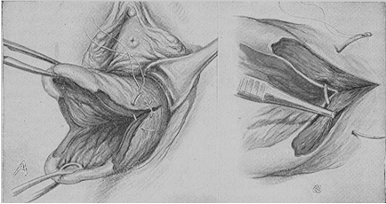
Treatment. The lacerated or ruptured cervix should be repaired by suturing. The sutures should be placed immediately after inspection of the cervix and discovery of lesions. The cervix should be pulled by the forceps toward the introitus and moved aside (in the direction opposite to the affected side). Suturing should begin from the upper angle of the rupture (the first suture should be placed slightly above the rupture) to the margins of the cervical os; the mucosa of the cervix should not be involved in the suture (Fig. 218).
If the upper edge of laceration is difficult to identify, the first suture should be applied slightly below, and the ends of the ligature should be pulled down; the upper angle of the wound will thus be revealed and become accessible for suturing. Catgut should be used to correct cervical lacerations and ruptures. The ends of ligatures should be cut off.

Fig. 218. Suturing of the cervix
Rupture of the Uterus. The disruption of integrity of the uterine walls is known as rupture of the uterus. If all layers of the uterus, i.e. endometrium, myometrium, and peritoneum break, the rupture is complete. The cavity of a completely ruptured uterus opens into the abdominal cavity. If only endometrium and myometrium are involved, the rupture is incomplete. A complete (penetrating) rupture of the uterus occurs more frequently than an incomplete one.
A comparatively thin lower uterine segment usually ruptures, but the upper segment and even the fundus can rupture as well. The rupture may occur along the line of the cervix attachment to the vaginal fornices; this is actually the separation of the uterus from the vaginal fornices.
Spontaneous and traumatic ruptures of the uterus are distinguished. A spontaneous rupture occurs without any extraneous cause, while a traumatic rupture is mainly due to improper operative intervention.
The rupture of the uterus is one of the most dangerous complications of labor. Even with modern obstetrical knowledge the rupture of the uterus not infrequently results in death of the mother and intrauterine fetus. The danger depends on acute loss of blood and shock. Blood is lost from the vessels which break together with the uterine wall. The larger the disrupted vessels, the stronger the hemorrhage. Another source of bleeding is the vessels of the placental attachment site. The placenta usually separates at rupture of the uterus and the vessels of the uterine wall at the site of placental separation start bleeding.
The hemorrhage accompanying the rupture of the uterus may be very profuse. The picture of acute anemia is aggravated by traumatic shock which usually occurs at rupture of the uterus, a complete rupture in particular. The shock develops on a background of strong irritation of the nerve endings of the ruptured uterus (especially of its peritoneal coat). The fetus is often extruded from the ruptured uterus into the abdominal cavity where it affects other abdominal organs increasing the danger of developing shock. The fetus would usually die very rapidly because of the placental separation.
The uterus may rupture due to a variety of causes. In the past century (1875) Bandl created a mechanical theory of uterine rupture. In his opinion, which was supported by many other obstetricians, the rupture occurs due to disproportion between the presenting part and the maternal pelvis. This would be usually explained by contracted pelvis, malpresentation (brow, posterior face presentation), a pathological asynclitism, large (gigantic) fetus, hydrocephaly, or transverse and oblique presentations of the fetus.
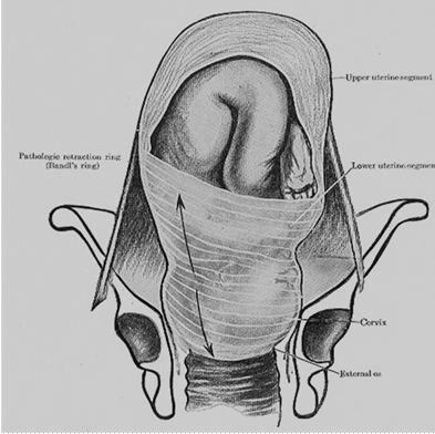
Violent uterine contractions develop in obstructed labor; the upper uterine segment contracts even more intensely, and the fetus is propelled gradually toward the lower uterine segment where the walls are thinned by distension. The contraction ring (at the junction of the lower and upper uterine segments) rises to higher levels to reach the navel (the ring may also be oblique). As the contractions persist, the lower uterine segment becomes overdistended and ruptures (Fig. 219)

Fig. 219. Excessive thinning of lower uterine segment
Verbov suggested another theory, according to which an intact uterus never ruptures and the rupture may only develop in the presence of pathological changes in the uterine wall which are responsible for inadequacy of the myometrium. The changes that predispose to the rupture of the uterus are scars resulting from previous cesarean section or other operations on the uterus (enucleation of a myomatous node), injuries associated with previous abortions, degenerative and inflammatory processes in the past history, infantilism, and other abnormalities.
According to modern views, the rupture of the uterus may result from both pathological changes in the uterine wall and mechanical factors. The uterus is more likely to rupture in cases with combination of these defects, i.e. in simultaneous action of several pathological factors (pathologies in the uterus concurrent with obstructed labor).
The uterus usually ruptures in multiparae and very seldom in young primiparae.
The rupture usually occurs during the expulsive stage of labor when obstacles to the progress of the fetus through the birth canal are encountered. In the presence of pathological changes in the uterine wall (scar tissue, inflammatory and degenerative processes), the uterus may rupture during the 1st stage or even at the very beginning of labor. Cases of uterine rupture during pregnancy are also known (due to pathological changes in the uterine wall).
Classification of uterine ruptures. Depending on time of occurrence:
· Rupture of the uterus during pregnancy
· Rupture of the uterus during labor
· Depending on pathogenesis:
· Spontaneous rupture: a) mechanical (due to disproportion between the presenting part and the maternal pelvis); b) histopathologic (due to pathological changes in the uterine wall); c) combined (due to both pathological changes in the uterine wall and mechanical factors).
· Violent (forcible) rupture of the uterus: a) traumatic (due to external effect during labor); b) combined (due to overdistended uterus and external effect).
· Depending on clinical picture:
· Threatened uterine rupture
· Complete uterine rupture
· Depending on damage:
· Fissure
· Incomplete
· Complete
· Depending on localization:
· Rupture of the fundus
· Rupture of the corpus
· Rupure of the lower segment
· Separation of the uterus from the vaginal fornices
Threatened Rupture of the Uterus. The clinical picture of threatened rupture is very specific in obstructed labor (contracted pelvis, malpresentation of the fetus, large fetus, etc.) The clinical picture of threatened rupture associated with the presence of mechanical obstacles to the expulsion of the fetus is as follows:
· Labor is violent; the contractions become convulsive in character.
· The lower uterine segment is overdistended, thinned, and painful on palpation.
· The contraction ring stands high (to reach the navel). Its position is oblique.
· The round ligaments of the uterus are very strained and painful.
· The margins of the cervical os become edematous (due to compression). The edema extends to the vagina and perineum.
· Urination becomes difficult due to compression of the bladder and the urethra between the bony pelvis and the fetal head.
· Traces of blood appear in the vaginal discharge to indicate the beginning of tissue injury.
· The parturient is excited, restless (she cries, tosses in bed, grasps the abdomen) and complains of sharp pain.
The clinical picture of threatened rupture due to a scarred uterus (scar on the uterine wall after previous operations) is less distinct, and differs in absence of precipitate labor: the contractions are frequent and painful but not very forceful. Other symptoms, such as overdistension, tenderness of the lower uterine segment, edematous cervix, vagina and external genitalia, disordered urination, etc., are also present but are less distinct than in a threatened rupture due to mechanical cause. In the presence of scars resulting from previous cesarean section the clinical symptoms of threatened rupture are the following: tenderness of the scar during contractions and on palpation, thinning of the scar (niche symptom). Threatened rupture of scarred uterus may be predicted with US of the uterus by evaluation of irregularity of uterine wall, niche appearance on the scar area.
If appropriate medical aid is not rendered, the uterus will inevitably rupture.
Threatened Uterus Rupture Management. When signs of threatened rupture develop the uterine contractions should be discontinued or lessened, and abdominal cesarean section should be performed on the patient.
Ether (or other type of inhalation narcosis) should be given in order to stop or lesson uterine contractions.
The woman should be delivered very carefully with deep anesthesia. If the fetus is alive and there are no signs of infection, a cesarean section should be made. A dead fetus should be dissected and removed by parts. Version of the fetus or application of obstetrician forceps is contraindicated; these operations, or even an attempt to perform them, will inevitably result in the rupture of the uterus.
A Complete Uterine Rupture. A complete rupture is characterized by the following symptoms:
· An extremely severe and acute pain (knife-like pain) in the abdomen is felt by the woman at the moment of rupture.
· Uterine contractions discontinue immediately following the rupture.
· The condition of the patient is sharply aggravated due to increasing anemia and shock. The skin and the visible mucosa become pallid, the face becomes pointed, the pulse is accelerated and weak, the arterial pressure drops. Nausea and vomiting often occur.
· When the uterus disrupts, part or the whole fetus is extruded into the abdominal cavity. Separate parts of the fetus become now directly palpable through the abdominal wall; the presenting part, which was previously engaged, moves upwards and becomes movable. The fetal heart tones are not heard. The contracted uterine body can be palpated by side of the fetus.
· External bleeding is usually not pronounced and sometimes even insignificant. The blood fills the abdominal cavity (a hematoma of the pelvic connective tissue develops in incomplete rupture of the uterus).
In the presence of pathological changes in the uterine wall (for example, in case of scar after previous operation), the rupture may occur because of gradual separation of the tissue. Acute and sudden pain may therefore be absent and contractions do not stop abruptly but lessen gradually. All other signs of the uterus rupture are obvious.
Management of Complete Uterine Rupture. If the uterus has already ruptured, a laparotomy should be performed immediately. The fetus, the placenta and the blood should be removed from the abdominal cavity. The uterus should then be extirpated. In some cases the uterus is repaired by suturing (in young women, if only little time has passed since the uterus was ruptured, and in the absence of infection). Measures to prevent blood loss and shock should be taken during the operation and immediately after it. Blood transfusion, intravenous infusions of physiological saline solution, plasma and albuminous solutions, cardiac preparations should be used.
Prophylaxis of the Uterine Rupture. It consists in adequate organization of obstetrical aid. Thorough observation of all pregnant women is very important in this respect. All women in whom the rupture of the uterus is likely to occur should be given special care. The factors predisposing to the uterine rupture are contracted pelvis, malpresentation of the fetus, post-term pregnancy, large fetus, flaccid abdominal wall and uterus in multiparae, unfavourable obstetrical anamnesis (pathological previous labor, complicated abortions, puerperal and postabortal inflammatory diseases), previous cesarean section and other operations on the uterus.
All women in whom the predisposing factors have been revealed should be hospitalized two or three weeks prior to anticipated labor. Labor should be conducted in the presence of a physician. The parturient should be observed thoroughly during labor so that the signs of threatened rupture are not overlooked.
Date added: 2022-12-25; views: 1049;
