Excessive Labor Pains (Hyperactivity of the Uterus). Justo Minor Pelvis
Excessive forceful contractions occur less frequently than uterine inertia. Violent and painful uterine contractions at short intervals develop in women with easily excitable nervous system, in patient with some neuroendocrine disorders. Violent contractions may develop in the presence of contracted pelvis, large fetus, malpresentations to the propulsion of the fetus through the birth canal. Under the impact of convulsive contractions the uterus may rupture. In the absence of cephalo-pelvic disproportion the excessively forceful contractions may result in precipitate labor, which lasts only from one to three hours and therefore often occurs out of hospital (at home, on the way to the hospital).
Clinical features are: significant, intensive pains with very short intervals between them. There are 5 contractions per 10 minutes. The uterus can not relax between contractions, the patient cries, is very irritable, intrauterine fetal hypoxia occurs due to the diminished placental circulation.
Complications of excessive labor pains are:
· maternal injuries (rupture of the cervix, vaginal walls, perineum, uterus, etc.)
· hemorrhages in the 2nd and 3rd stages of labor, in early puerperium
· fetal injuries (intracranial injuries), fetal asphyxia.
Treatment. Spasmolytics, adequate analgesia (2 ml of 1% promedol, 5 ml of baralgin intravenously), peridural anaesthesia may be used. Medicamental tocolysis is usually prescribed: partusisten 5 mg in 500 ml of 5% dextrose intravenously droppingly. The starting rate is 6-8 drops/minute. Every 10 minutes the rate of drops must be increased by 6-8 drops till the uterine activity becomes optimal. Due to the cardiac side-effects of these drugs this infusion should be done under careful measurement of pulse rate and arterial blood pressure. Increasing of the pulse rate to 120 beats/min means the availability of side-effect, and in such cases the rate of drops cannot be increased. If there is no effect of treatment cesarean section is indicated to prevent severe maternal and fetal complications.
Uncoordinated Uterine Activity (Uncoordinated Labor Pains). This variety usually appears in an active stage of labor. Uterine contractions may begin in its lower but not the upper segment. They may begin from any other part of the uterus but not from the upper segment as normally. Sometimes every part of the uterus contracts in its own rate and rhythm. Uncoordinated contractions are very painful and ineffective, the cervix dilates at a slow rate and the progress of the presenting part is delayed.
Spastic lower segment: fundal dominance is lacking and there is reversed polarity. Inadequate relaxation in between contractions is responsible for painful and non-effective labor.
Constriction ring: there are localized spastic contractions of a ring of circular muscle fibres of the uterus. It is usually situated at the junction of the upper and lower segment around a constricted part of the fetus, usually around the neck in cephalic presentation. The patient feels significant pain, the progress of presenting part is absent, intrauterine fetal asphyxia occurs due to the change of placental circulation. The uterus may rupture.
Cervical dystocia: the normal pattern of uterine contraction is maintained but the external os fails to dilate. It may be due to the presence of excessive fibrous tissue or spasm of circular muscle fibres surrounding the os. The cervix becomes very much thinned out and well applied to the head. Initially, the uterine contraction remains good but ultimately becomes ineffective. On occasion, edema of the anterior lip may occur and delivery may be accomplished by avulsion of the anterior lip or by annular detachment of the cervix.
Uterine tetanus: a pronounced retraction occurs involving the whole of the uterus up to the level of the internal os. Thus, there is no physiological differentiation of the active upper segment and the passive lower segment of the uterus. Every part of the uterus contracts in its own rhythm and intensity. There isn’t any possibility for uterine relaxation, so fetal asphyxia may occur very quickly. The rupture of the uterus may also occur; shock due to significant pains may be too.
Treatment. The main principles of treatment of uncoordanated labor pains are: sedative therapy, spasmolytics, anaesthesia of labor (peridural is better), medicamental tocolysis. In cases of ineffective therapy cesarean section must be done.
Justo Minor Pelvis. Justo minor pelvis has the same shape as a normal one while all diameters are shortened. For example, typical sizes are: interspinous diameter – 24 cm, intercristal diameter – 26 cm, intertrochanteric diameter – 29 cm, external conjugate – 18 cm, etc. (Fig. 198).
The rhomboid is shortened both in vertical and horizontal axis.

Fig. 198. Justo-minor pelvis
Mechanism of Labor in Justo Minor Pelvis. The 1st moment is a strong flexion of the head. The sagittal suture aligns with one of oblique diameters of the pelvic inlet. The biparietal diameter of the head passes the oblique diameter of the pelvis which is longer than the anteroposterior one. The engaging diameter of the head is a small oblique (suboccipitobregmatic) diameter. The leading point (denominator) is a small (posterior) fontanelle. A strongly flexed head descends into the pelvic cavity and then performs the same movements as in normal mechanism of labor: the internal rotation, extension, external restitution.
Because of narrow pubic angle, the posterior cranial fossa comes in direct contact with the symphysis as the head passes the pelvic outlet plane. The head is therefore displaced toward the perineum to a greater extent than in normal pelvis, the perineal tissues undergo greater extension and deep laceration of the perineum may occur. But all movements are comparatively slow and require much effort of the parturient.
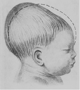
The head of the delivered fetus is elongated in the direction of the occiput (dolichocephalic shape) and a considerable swelling is formed in the region of the posterior fontanelle (Fig. 199).

Fig. 199. Dolicho-cephalic shape of the head
Transverse Contracted Pelvis. One or more transverse diameters may be shortened in transverse contracted pelvis. Transverse contracted pelves are usually elongated in anteroposterior direction. There are a lot of varieties of transverse contracted pelvis, which depend on the shortened diameter, degree of shortening, etc. For example, sizes of transverse contracted pelvis are: interspinous diameter – 23 cm, intercristal diameter – 26 cm, intertrochanteric diameter – 28 cm, external conjugate – 21 cm. Diagonal and true conjugate may also be normal or even bigger than in normal pelvis (Fig.200). The rhomboid of Michaelis is elongated in its vertical axis so that its upper and lower angles are acute, and lateral ones are obtuse.

Fig. 200. Transverse contracted pelvis
Mechanism of Labor in Transverse Contracted Pelvis. The 1st moment is flexion of the head. The sagittal suture aligns with one of oblique or anteroposterior diameter of the pelvic inlet (depending on shortening of transverse diameter), the engaging diameter of the head is suboccipitobregmatic (small oblique), the denominator (leading point) is a small (posterior) fontanelle. The head enters the pelvic cavity and sometimes even reaches the pelvic floor without any rotation. This condition is termed as high anteroposterior situation of the sagittal suture. Further the head is delivered as in occiput presentation.
Date added: 2022-12-25; views: 833;
