The organization of hereditary material of non-cellular forms, prokaryotes and eukaryotes
The non-cellular life forms are viruses and bacteriaphages (pic 2.5). Viruses - are non-cellular life forms, which are able to enter to special live cells and reproduce itself only inside of these cells. Bacteriaphages are viruses of bacteria. There is only one type of nucleic acid in the viruses (DNA or RNA). By this, viruses can be divided to RNA-containing and DNA-containing. A nucleic acid serves as storage of hereditary information.
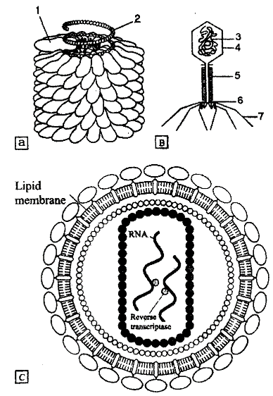
Pic. 2.5. The scheme of non-cellular forms Structure. A-tobacco mosaic virus (1-protein coat, 2 - RNA molecule); В - bacteriaphag T4 (3 - DNA, 4 - head, 5 - tail, 6 - base plate, 7 - tail fibers); C - HIV
(human immunodeficiency virus) (K. Swenson, P. Webster, 1980 and R. Gallo 1987)
All viruses are divided into simple or complex. The simple viruses consist of nucleic acids and protein coat (capsid). The complex viruses may also have lipoprotein membrane, carbohydrates and non-structural proteins. The size of viruses may vary from 15 to 2000 nanometers. The molecular weight of viruses DNA is around 200x10*6 Daltons and viruses RNA is from 10*6 to 15x10*6 Daltons. The nucleic acids vary in shape. There may be single strand RNA and double strand DNA as well as double stranded RNA and single stranded DNA.
The RNAs as usual are linear. Some viruses may have a set of RNA fragments, each carrying part of necessary information for virus reproduction.
The genetic material of bacteria is organized as a single, circular molecule of DNA (pic 2.6). The E.coli has a DNA of 1mm long. It has 4x10*6 nucleotide pairs, making around 4000 genes. The most of prokaryote DNA (95%) is actively transcribed in any moment of time. There are no histons providing nucleosome organization of genetic material. The DNA molecule of prokaryotes folds in a form of loops. Then it binds some histons to form nucleotide. The nucleotide is less stable as chromatin of eukaryotes.
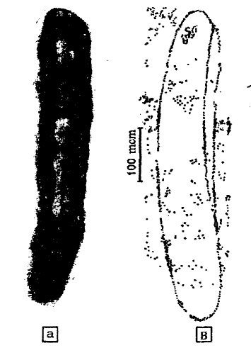
Pic. 2.5. Escherichia coli. А - general view (lighter part present DNA); В - autoradiogram of circle chromosome. DNA is marked by tritium. On a left side, die replication beginning is visible. (S.M. Gershenzon 1979)
The genetic material of eukaryotes in interphase nucleus is presented by chromatin. When cell divides by mitosis, the chromatin is spiralized to chromosomes. Besides DNA, chromatin contains many different proteins. Most of them are histons. The histons are proteins with positive charge and molecular weight 10000-20000 Daltons. They have 5 classes: Hl, H2A, H2B, H3, and H4. The HI contain a lot of lysine, H3 - arginine, H4 - arginine and glycin. The others, so called non-histon proteins, are in a small amount. According to common point of view, chromatin is presented by spiraling strings. There are following levels of chromatin folding (pic 2.7).
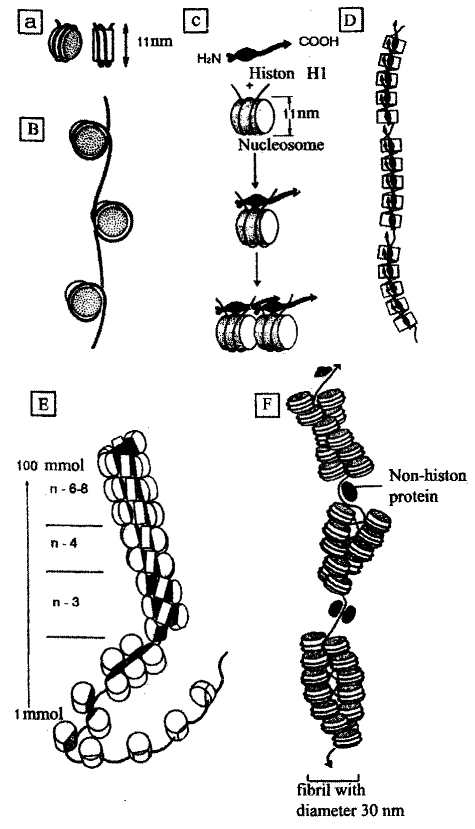
Pic. 2.7. Molecular organization of chromosome: a - free nucleosome; b * nucleosome string; c - nucleosome connection by histon; d - chain of nucleosome groups divided by free DNA regions; e - dependence of DNA spiralization from NaCl concentration (100 mmol NaCl correspond to 6*8 nucleosomes on one turn of helix, the lower concentration corresponds to lower nucleosomes number); f - interphase chromonemm (V.N. Yarygin, 1997 and F. Fogel, A. Motulsky, 1989)
The nucleosome string. This level of chromatin organization is provided by four types of histon proteins: H2A, H2B, H3, H4. They form a proteins bodies, which look like a puck, - the cores. The cores are formed from 8 histons (2 of each type). The DNA molecule spirally turns over proteins core. One core is covered by 146 nucleotide pairs of DNA. The cores are connected with each other by linker DNA. The linker may be 15 to 100 nucleotides long.
It depends on cell type. In experiments in vivo, it is shown that structure of nucleosome string depends on the NaCl concentration. So, if the concentration is 100 mmol, one spiral turn has 7-8 nucleosomes. If the concentration decreasing, each spiral turn has 3-4 nucleosomes. With help of nucleosome, chromatin organization the double helix of DNA with diameter 2nm and average length 5 cm achieve a diameter 1011 nm and length 2cm.
Chromatin fibril. Next chromatin folding is provided by H1 histon protein. It is bounded with linker DNA and is put nucleosomes close to each other. Such chromatin fibril, so called elementary febrile, has the diameter 20-30 nm and length 1.2mm.
Interphase chromonemm. This level of chromatin folding is provided by folding of chromatin fibrils to loops. The non-histon proteins take part in this process. They merge pointed regions making the loop with the fragments of chromatin fibril in it. The one loop contain from 20000 to 80000 nucleotide pairs. After such folding, interphase chromonemm has the diameter 100-200 nm.
The regions of interphase chromonemm undergoing further folding makes a structural blocks, which can be visible in the interphase nucleus as chromatin particles. There are euchromatin regions and heterochromatin regions, according to their functional activity. The euchromatin regions have a less tight folding because of active transcription processes. The heterochromatin regions have a tighter folding because of lack of transcription processes. There is constitutive and facultative heterochromatin.
The constitutive heterochromatin is in the telomere regions and regions near the centromere and along some internal fragments. It is believed constitutive heterochromatin to provide keeping of total nucleus shape, attaching chromatin to karyolemm, participating in chromosome recognition during meiosis, making an intervals between genes.
The facultative heterochromatin has information. It contains genes and may be changed to euchromatin. The example of facultative chromatin is a sex chromatin body, which is in the cells of organisms with homogametic sex. Also facultative chromatin formation occur during processes of cell differentiation, serves as a mechanism of switching off activity of several genes which is not necessary in the cell of such specialization.
Metaphase chromosome. In the beginning of mitosis, chromatin condenses to chromosomes. Chromosomes become visible. The mitotic superspiralization makes process of chromosome movement easier.
The chromosome DNA consist of more than 10*8 nucleotide pairs, which form information blocks - genes, placed linearly. They represent 25% of total DNA.
The gene is a functional unit of DNA, containing information for protein or RNA synthesis. There are spacers between genes. It is non-informative regions of DNA of different length. The excessive genes are presented by a large amount of identical copies, for example genes for tRNA and rRNA. In the DNA, there are the sequences of the same nucleotides.
They may be moderate and highly repeating. The moderate repeating sequences are 300 nucleotide pairs of length and usually they are the spacers and excessive genes. The highly repeating sequences makes constitutive heterochromatin. There is around 75% of chromatin non-participating in transcription. This is highly repeating sequences and nontranscribed spacers.
Date added: 2022-12-30; views: 811;