The endomitosis and polyteny. The amitosis. The cell proliferation
The endomitosis - is one of mitosis type. During endomitosis occur only chromosome replication but it is not following by cell division. As result of this, the chromosome number in a cell is multiplied, sometimes in more than ten times.
The endomitosis occurs in intensively working cells: in tissues of nematodes, insects, in some plants. It is assumed that endomitosis appeared in evolution as a variant of mitosis.
The polyteny - is chromonemms reproducing in chromosomes without increasing chromosome number. The number of them might be increased in many times (in 1000 and more). The chromosomes grow until giant sizes because of DNA amount increasing. The polyteny was firstly described by A. Balbiani in 1881. The all phases of mitotic cycle are lost except chromonemm reproduction. If we stain these chromosomes, we can see dark strips (disks) across the chromosome They appear because of irregular chromosome spiralization (pic 3.19).
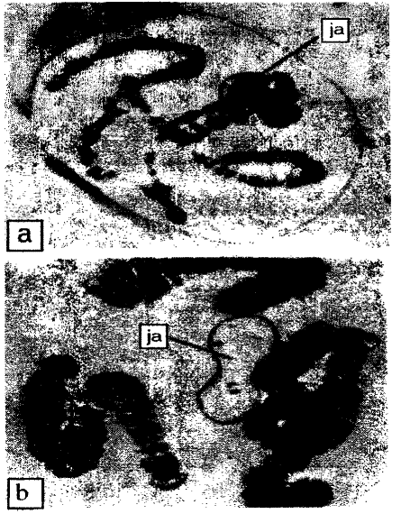
Pic. 3.19. The polytenic chromosomes in nucleuses of salivary gland cells of Drosophila a - chromatin in the nucleus; b - enlarged chromosomes with nucleolus (ja) (by O.Necasu et al., 1969)
The polyteny occurs in some insects, infusoris and some plants. In a drosophila, salivary gland cells the ploidity of chromosomes reach 1024. The polyteny is used for chromosome mapping and revealing chromosome changes.
The amitosis. The amitosis (from Greek, a- negative, mitos - thread) or direct division of cell is nucleus division without chromosome spiralization and assembling mitotic apparatus. In 1841 R. Remark was first to describe amitosis. During direct division, firstly, nucleolus is divided into two parts, and then such division occurs with nucleus and cytoplasm (pic 3.20).
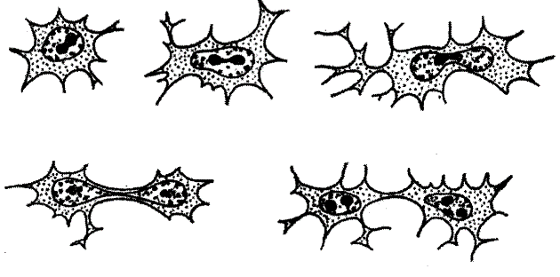
Pic. 3.20. The amitosis: stages of division of synovial mouse cell (by P.B. Gofinan-Kadoshnikov, 1966)
The nucleus may be divided into two equal parts (equal amitosis) or into two non equal parts (non equal amitosis), or into several parts (fragmentation, plasmodium shysogony). Sometimes the cytoplasm is not divided and then cells having many nucleuses are created (amitosis without cytotomy).
There are several factors that may lead to amitosis. According to these factors, amitosis may be divided to three types: generative, reactive and degenerative (Zhilkin L.N, 1966).
The generative amitosis may occur while highly spetialized cell division. It is in infusoria while macronucleus division, in some mammalian cells (liver cells, epidermis cells).
The reactive amitosis may occur while cell undergo to some harmful impacts or during metabolism disbalancing (fasting, tissue denervation, disturbances in nucleic acids exchange).As usual it has no cytotomy. It leads to multinuclear cell formation. Possibly, it may be considered as compensatory organism reaction resulting in increasing metabolic surface between nucleus and cytoplasm.
The degenerative amitosis may occur only in aging cells. It is presented by nucleus fragmentation and it has no any connection to cell reproduction. The appearance of degenerative amitosis form is a sigh of necrobiotic processes.
The cell proliferation. The proliferation is an increasing of cell number by mitosis, leading to tissue growth. Contemporary, cells of animal tissues may be divided into three main groups: labile, stable and static.
Labile cells are the cells, which are able to renew itself fast and easy during organism life (blood cells, epithelial cells, cells of alimentary channel mucosa).
Stable cells are the cell of such organs as liver, pancreas, salivary glands etc. They have a limited ability to reproduction. This ability appears only during reparation of damaged organ.
Static cells are the cells of myocardium and nervous tissue. They are not subject to division or subject to division in extraordinary conditions.
The process of wound healing connected with cell division. The value of proliferation is determined by tissue ability to division. None wound may be healed without cell division. And operating surgeon has to consider the ability of the cell and tissues to reproducing (proliferation).
The mechanisms providing cell division. A pioneer in studying of reasons and controlling factors, which are responsible for cell division, was A.G. Gurvich (1874-1954). He pushed forward the hypothesis about influence of special mitotic rays on division process. L. Y. Bliaher (1954) and I. A. Utkin (1959) showed an important role of neurohumoral regulation of mitotic activity. It was stated that epinephrine secretion suppress mitotic activity, but thyroid hormones activate mitosis. Removal of the adrenal gland leads to switching off effect of mitosis suppression. It was stated that there are many reasons leading to mitotic divisions.
It was proved that all synthetic processes in a cell preparing to division are controlled by its genetic material. The genes controlling this process are in different chromosomes. F. Jacob and J. Monod (1961) suggested a hypothesis of gene activity regulation in prokaryotes. This hypothesis may be used today and for explanation of eukaryotes gene activity.
The cell biology is very important for understanding ontogenetic life organization level. It is still needed to make completely examination of all structural cell components, to understand reasons of determination, differentiation and synchronization of cellular reproduction, to clear mechanisms of controlling, regulation and integration of cell and its organelles. Thus, answering this question is important for.
Date added: 2022-12-30; views: 998;