The gametogenesis. The features of gametes structure
The gametogenesis is a process of sex cell formation. All cells of a body, somatic and reproductive, have their origin from embryonic cells. During embryonal development group of cells separates from others. And after several divisions they form gonial cells - gonia. At the beginning they are the same, but later they subject to differentiation. In a male organism they differentiate to spermatogonia, in a female organism to oogonia. The gametogenesis has four periods: reproduction, growth, maturation and formation (pic 4.4).
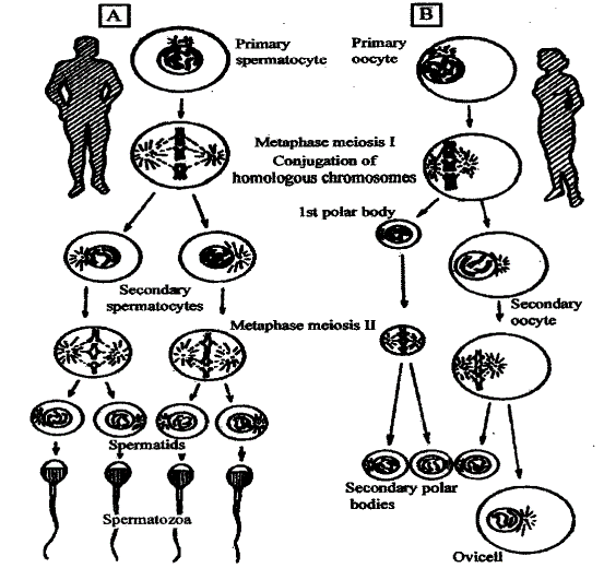
Pic. 4.4. The spermatozoa (A) and ovicells (B) formation in human (by N. P. Dubinin, 1976)
The spermatogenesis. During a first period (period of reproduction) cells of sexual germ are presented by spermatogonia. It is small round shape cells with a small amount of cytoplasm, dividing very actively. They are subject to division almost all life long, from childhood to eldely. At puberty onset, the part of spermatogonia stops their division and they are changed to spermatozoa.
A growth period is characterized by reproduction termination and spermatogonia are changed to primary spermatocyte. They grow, increasing their size in four times. They lie in semineferous tubules closer to duct. During maturation period a meiosis division is performed. As result of this primary spermatocytes are changed to secondary spermatocytes and then to spermatids. Secondary spermatocytes are in two times less in volume than primary spermatocytes.
However, spermatids are in four times less in volume than primary spermatocytes. They lie closer to duct lumen than primary spermatocytes. During period of formation, the spermatids are changed to spermatozoids
The oogenesis. Oogonia have a reproduction period only during embryonic development. At the end of this period, oogonia stop reproducing and are changed to primary oocyte. They are preserved in ovarium until puberty. At the puberty onset, the growth period starts in selected oocytes. It may be distinguished “small growth”, nucleus and cytoplasm volume increasing, and “large growth”, accumulation of yolk inclusions (proteins, fats, fats-like substances). There is a lot of yolk in amphibia, reptilia and birds ova, but there are a few yolks in lancelets, mammalian and human ova.
The nucleus is changed to badly stained vesicle. Many animals loose centrosome. During maturation period two irregular meiosis divisions occur. Primary oocyte gives up the secondary oocyte and the first polar body. Then secondary oocyte gives up second polar body and after that, it becomes mature ovicell. The first polar body may be divided to two polar bodies. This irregular division may be explained by expediency of yolk and cytoplasm preservation for ovicell.
Thus, the main differences of oogenesis from spermatogenesis may be concluded in following:
1) the reproduction period of oogonia is terminated after birth;
2) The oogenesis growth period is longer and have subdivisions to “small growth” and “large growth”. Oocyte becomes bigger than spermatocyte;
3) Primary oocyte may give only one full gamete, whereas spermatocyte gives four;
4). In oogenesis period of formation is almost absent.
The sex cells, which were formed in gametogenesis, have a following structure.
Ovicells - are oval, big, immobile cell which are in hundreds or even millions times bigger than spermatozoa. Many animals have ovicell without centrosome, unable to be divided.
There are several ovum types according to yolk amount and distribution. Isolecitinal ovicells (primary and secondary) have a satisfactory amount equally distributed yolk, with nucleus in central of a cell. Polylecitinal ovicells (centrolecitinal andtelolecitinal) have excessive amount ofyolk. Alecitinal ovicells have very little equally distributed yolk. The ovicells of mammalians and flat- worms are alecitinal. However, some researchers consider mammalian ovicell to be isolecitinal as many mollusks, lanceolate ovicells are. In telolecitinal ovicell yolk is distributed irregular.
It is very little yolk near animal pole of such ovicell. A large amount of yolk is on vegetative pole of ovicell. The examples of such ovicells are ovicells of amphibia, reptilia and birds. Centrolecitinal ovicell has a large amount equally distributed yolk. However, near cell membrane there is a cytoplasm layer without yolk. The nucleus of such cells also is surrounded by the same cytoplasm layer. Arthropods have ovicells of this structure.
The ovicell is protected by coats (pic 4.6a). There are primary coat produced by ovicell itself, secondary coat, produced by follicular cells, tertiary coats that surround ovicell while it moves in uterine tube.
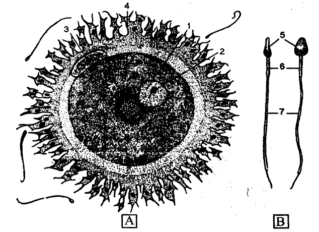
Pic. 4.6. The human ovicell (A) and spermatozoon (B) (x2000); I - nucleus, 2 - nucleolus, 3 - polar body, 4 - corona radiate, 5 - head, 6 - neck, 7 - tail (by K. Villy, V. Detier, 1971)
All animals have primary coat. It also called yolk coat. Humans and mammalians have it as an internal part of dense coat. The external part of dense coat is produced by follicular cell and it is secondary coat. Microvilia of ovum enter to the dense coat from inside and microvilia of follicular cells enter from outside. On a high power magnification, it is looked striated and that why called radiated coat “corona radiata” or shining coat “zona pellucida”. The dense coat contains primary and secondary coats.
Tertiary coats are well developed in reptilians, birds, amphibians and cartilaginous fishes. These coats have no cellular structure. They are produced by uterine tube mucosa to defend ovum from different harmful influences. Those animals that live on a land use such coat for water and food storage for embryo.
Spermatozoa - are a small, mobile cell with nutritive substances storage reduced to minimum. A sperm has head, neck and tail. In the head there is a nucleus surrounded by thin cytoplasm layer. There is acrosome on a top of the head. Acrosome is derived from complex Golgi. It is consist of compact mass and membrane. It contains active substances facilitating ovum coats penetration by sperm. There are two centrioles, proximal and distal in a sperm neck. Distal centriole forms axis thread of a tail. Proximal one takes part in cell division after fertilization. Tail is an organ of movement. The core of a tail is axial thread. It is surrounded by mitochondria (in a main part) providing energy for movement.
A sperm brings centrosome to ovicell while fertilization. The cytoplasm of sperm head has liquid-crystal state, which defend it from harmful environmental influences. Sperm has high nucleus/cytoplasm ratio corresponding to it main task — to bring genetic material to ovicell. Acrosome enzymes help to dissolve ovicell coat.
Date added: 2022-12-30; views: 943;