The features of human spermatogenesis and oogenesis. Their regulation by hormones
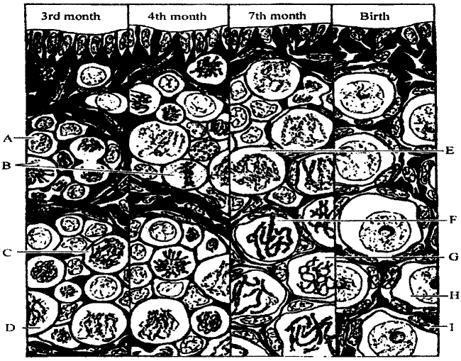
When an embryo has reached a size 20 mm, specific sex features of female become evident. The primary sex cells, incorporated in gonad germ, proliferate and are subject to differentiation to ovogonia in ovariums of embryo at 2nd month of development. At the end of 3rd month in deep layer of female gonad, it may be distinguished a differentiated oocytes in prophase of meiosis L At 7th month, the histological differentiation of ovarium is very active.
So, at 9 month there are 200000-400000 oocytes in an each embryo ovarium. Some investigators state that there are about 1 million oocytes (pic 5.1). Oocytes are surrounded by follicular cell monolayer and form primary follicule. After birth, oocytes are preserved until puberty in diplonemm of meiosis prophase I. When puberty has been reached, the oocytes continue their meiosis. First meiosis division has done before ovulation (liberating of ovicell from follicule).

Pic. 5.1. The oogenesis in human female embryo: A - interphase; В - metaphase; C - anaphase; D - leptonemm; E - zygonemm; F - pahynemm; G - diplonemm; H - diakinesis; I - oocyte surrounding cell (by S.Ohno, 1962)
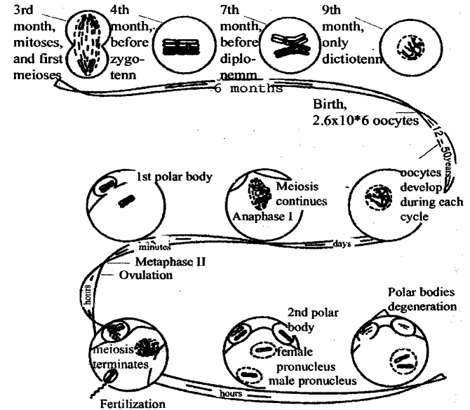
This division is very unequal. Secondary oocyte gets the most of cytoplasm, whereas a polar body gets a minimum. The second meiotic division does not occur until fertilization and result in production of second polar body and a single haploid egg nucleus. Both cells move into fallopian tube, where polar bodies are destroyed liberating nucleus substance into surrounding ovum environment (pic 5.2).

Pic. 5.2. The scheme of vogenesis in female organism (by KBresch, M.Hausman, 1972)
Now it is apparent that regular follicule growth, ovulation and regression is regulated by follicle-stimulating hormone (FSH) and luteinizing hormone (LH) of pituitary gland. Growing follicules produce estrogens (estradiol) which may act on pituitary hormone production. It is stated that follicule growth mostly depends on FSH, but ovicell maturation and ovulation mostly depends on LH. The hormone mechanism integrates two different, evolutionary unconnected processes as follicule growth and ovulation.
This allows providing fully differentiated gametes for fertilization. To trigger meiotic division it is necessary to have a small amount of LH. But to perform ovulation we need to have a peak of LH concentration. So, in some cases, ovicell has done meiotic division, but LH concentration isn’t enough to perform ovulation. At this situation an intrafollicular aging of ovum occurs. The properties of ooplasm are changed, which is mainly concerned for cortical layer and for spindle apparatus.
This cause an ovum death, loosing fertilization ability, or formation of zygote with unbalanced chromosome set. This resulting in embryo death and formation of embryo with chromosome defects (such as Dawn syndrome). It is believed that intrafollicular ovicell aging is connected with seasonal disturbances in neurohomonal regulation pattern.
A male primary sex cell is subject to differentiation to spermatogonia when a male embryo has reached a size 15 mm. A specific sex signs formation in a male embryo starts earlier than in a female embryo. A period of primary spermatogonia formation is very short. During this period, many mitotic abnormalities occur, such as failures in chromosomes moving.
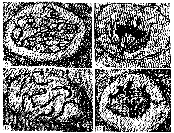
Many cells die at this stage. The process of male’s gametes formation continues throughout all life. A process of sperm formation takes about 70 days. Each day 10*7 spermatozoa are produced per 1 gram of testis weight. The epithelium of semineferous tubules consist of external layer of germinative epithelial cells and six inner layers corresponding spermatozoa formation stages. The division of germinative cell gives a rise to many spermatogonia, which increase in size and become primary spermatocytes. Primary spermatocytes are subject to meiosis I forming secondary spermatocytes. They becomes spermatids after meiosis II (pic 5.3).

Pic. 5.3. The meiosis in human spermatogenesis: A - zygonemm (conjugation of homologous chromosomes): В - pahynemm; C - metaphase I; D – anaphase II (by B. Severinghause, 1942)
There are Sertoli cell in-between developing lines of cell. They perform nutrition for developing cells and they also secrete a fluid that helps spermatozoa to move inside of the tubules. In an inner layer, spermatozoa are formed from spermatids. A growth and reproduction of sperms is stimulated by follicle-stimulating hormone. A testosterone secretion is stimulated by luteinizing hormone. The testosterone is a main male androgenic hormone.
It stimulates development and maintenance of male primary and secondary sexual characteristics. To produce spermatozoa successfully it is necessary to have both testosterone and FSH. Whereas a development and maintenance of male secondary sexual characteristics requires only testosterone.
Date added: 2022-12-30; views: 862;
