The cell physiology. The mitosis and meiosis
One of the main biological properties of the cell as an elementary life system is its ability to self-reproduce. Cell reproduction provides organism growth, development and regeneration. The time between cell formation by mother cell division and it own division or death is called cell cycle. For cell of an undividing cell populations the cell cycle is time between cell formation by mother cell division and it own death.
The mitotic cycle is obvious component of cell cycle. The mitotic cycle is a time between two cell divisions and all processes that occur during this time. The mitotic cycle of growing population may be divided to two big periods: the period between divisions - an interphase, when cell grow, perform it function, and get preparePI to divide; and cell division - mitosis. There IS cell growth, DNA replication, duplication chromatid number, producing of mitotic spindle proteins, energy producing and storage during interphase.
The interphase may be divided to three periods.
Postmitotic or presynthetic period, period G1. During this period cells grow, produce RNA, proteins, store energy, but they don’t make DNA. In presynthetic period cell nucleus contain diploid chromosome number, each chromosome contain only one chromatid. Chromosomes are despirilized. If we mention that DNA amount in 23 chromosomes is C, so the DNA amount in G1 is 2C.
Synthetic period, S period. During this period DNA replication occur. Each chromosome receives second chromatid. As a result of this amount of DNA after S period is 4C and chromosome number is diploid, each chromosome contain 2 chromatids.
Postsynthetic or premitotic period, period G2. During this period there is producing mitotic apparatus proteins and producing and storing energy for further mitosis. The next step is mitosis. The initial signal of mitosis start is changing of nucleus/cytoplasm ratio.
The integrity of processes to prepare cell for development and mitotic division itself are mitotic cycle of a cell. If daughter cell immediately begins to prepare for next division, their mitotic cycle and cell cycle are the same. In other cases daughter cell are subject to differentiation and carry out different functions. Their cell cycle is finished by their death.
There are two types of cell divisions: indirect division (mitosis) and direct division (amitosis). The mitosis consists of mitosis itself, meiosis, endomitosis and polyteny. The amitosis is divided by shape (equal, non-equal, multiply, without citotomy) and by type (generative, reactive, degenerative).
The first to describe mitosis phases was I.D. Chistiakov in 1874. The detailed description of plant cell mitosis was made by E. Strassbourger (1876-1879) and animal cell mitosis by V. Fleming (1882).
The mitosis. The mitosis (from Greek “mitos” - thread) - is unique type of animal and plant cell division, during which cell pass a range following changes leading to two daughter cell formation with diploid chromosome number and full range of genes, which are necessary for all individual hereditary properties development. The mitosis is subdivided into five phases: prophase, prometaphase, metaphase, anaphase and telophase (pic 3.16).
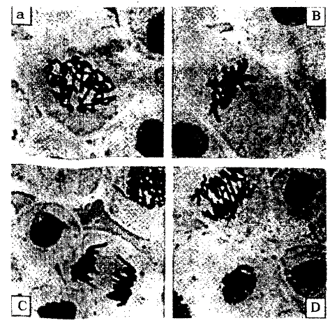
Pic. 3.16. The mitosis in cells of onion root: A - prophase; В - metaphase: C - late metaphase; D - early anaphase and late telophase (by O. Necasu et al., 1969)
The prophase. In a cell, incoming to division, chromosomes condensate and become visible by light microscope. In early prophase centriole divide into two parts and each part moves to opposite cell pole. At the same time the condensation process continues. It results in chromosome shortening and increasing chromosome width. There is diploid chromosome number in a nucleus.
Each chromosome consists of two chromatids, the DNA amount is 4C. Between centriols a radiate figure is formed. The nucleolus dissolves under the lysosomes action. The division spindle is made of two tubules types. The first one is polar, connecting both centriols, the second one is chromosomal, bounded to chromosome centromere.
The prometaphase. The cell cytoplasm has a small viscosity. Embedded in cytoplasm, chromosomes moves toward cell center. The nucleus coat is dissolved.
The metaphase. It begins when the pairs of sister chromatids align in the center of the cell. They are good visible, that’s why chromosome counting is performed at this stage. Each chromosome splits along itself on two chromatid. The nucleus characteristic is 2n - 2chromatids - 4C.
The anaphase. During this stage occurs chromatid movement toward cell poles. Such chromatid become a sister chromosomes. The spindle threads contract and pull chromosomes to cell poles. There are very active processes in cytoplasm, which is look like boiling fluid, while microphotographing. There are two chromosomes set at the end of movement on a cell poles. Each has diploid chromosome number, 2n, 1 chromatid, 2C DNA amount.
The telophase. The daughter chromosomes despiralize, loose good visible state. They are surrounded by new nucleus coat. The nucleolus is formed. The cell center looses its activity. The cytotomy (the cell cleavage) begins. The nucleus characteristics are 2n, 1 chromatid, 2C DNA amount.
The mitotic cycle duration is different. It may vary from several minutes to hundreds of hours. It is depends on tissue type, physiological organism state and environmental factors (temperature, light, chemicals etc.).
The meiosis. This type of division appeared as a special mitosis form providing sexual reproduction of organisms. As meiosis result, 4 haploid cells are formed from one somatic cell with diploid chromosome number. The meiosis has two following divisions: the first - reducing division, which decreases chromosome number in half (meiosis I), the second - equalizing division when a cell save their haploid chromosomes set (meiosis II) (pic 3.17). The most complicate is meiosis I. It has elongated prophase consisting of five stages.
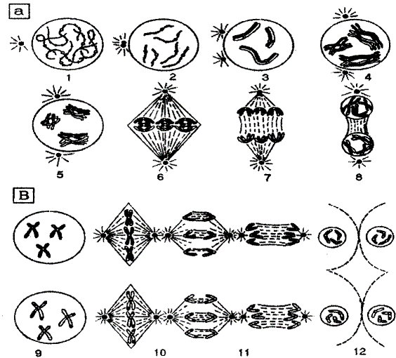
Pic. 3.17. The Meiosis scheme: A-meiosis I (I - leptomenn; 2 - zygonemm; 3 -pahynemm; 4-diplonemm; 5 -diakinesis; 6 - metaphase I; 7-anaphase I; 8-telophase I); В-meiosis II (9 - interphase; 10 - metaphase II; 11 - anaphase 11; 12-telophase II) (by D.G. Hamden, 1974)
The leptonemm. It is characterized by increasing nucleus volume. The diploid chromosome set becomes well visible. The chromosomes are thin, each containing two chromatids.
The zygonemm. There is chromosome conjugation. The homologous pairs of chromosomes line up side by side and then they exactly join, each gene located directly across from its corresponding sister on the homologous chromosome.
The pahynemm. It is very long. The conjugated chromosomes lie very tight to each other, forming bivalents. The bivalent consist of 4 chromatids. At this stage the crossing-over process occurs. The homologous chromosomes exchange some fragments that lead to genetic information exchange. It is one of combinating diversity mechanisms.
The diplonemm. The chromosomes start to coil. The chromosomes of bivalent begin to move apart. This movement starts from centromeres. The points at which portions of chromosomes have been exchanged can often be seen under the light microscope as an x-shape structure known as a chiasm.
The diakinesis. The chromosomes continue to coil. They become short and wide. The nucleus coat dissolves.
The metaphase I. The homologous chromosomes are by pair at the cell equator.
The anaphase I. The homologous chromosomes start to move toward cell poles.
The telophase I. The two cell containing haploid chromosome set, 2 chromatids and DNA amount 2C, are formed.
Between meiosis I and meiosis II is a short time period called interkinesis. During which chromosomes uncoils. The meiosis II occurs as a usual mitosis. The only differences are that there is a haploid chromosome set on equator in metaphase II and in anaphase II chromatids are moved to cell poles. In telophase II a cell containing haploid chromosome set, 1 chromatid and DNA amount 1C, are formed. Their destiny may be different: to be used for zygote formation or to die.
Date added: 2022-12-30; views: 749;