Chromosomes. The Stages of Mitosis
The Mendelian laws were more significant in 1900 than they were in 1866 because in the interim important new discoveries had been made concerning cells.
Those who observed cells during the eighteenth and early nineteenth centuries did not see much, even with improved microscopes. The cell was a virtually transparent body and so was the material within it. Consequently, it seemed a more-or-less featureless blob, and biologists had to be content to describe its over-all size and shape, and no more. Some occasionally made out a denser region (now called the "cell nucleus") near its center, but the first to recognize this as a regular feature of cells was the Scottish botanist, Robert Brown (1773-1858), who made this suggestion in 1831.
Seven years later, when Schleiden advanced the cell theory (see page 57), he attributed considerable importance to the cell nucleus. He felt that it was connected with cell reproduction and that new cells budded out of the nuclear surface. By 1846, Nageli was able to show that this was wrong. However, Schleiden's intuition did not lead him altogether astray; the nucleus was involved in cell reproduction. Knowledge concerning the details of this involvement, however, had to await some new technique for viewing the cell's interior.
The technique came by way of organic chemistry. Following the lead of Berthelot, organic chemists were rapidly learning how to prepare organic chemicals that did not exist in nature. Many of these were brightly colored and, indeed, the 1850s saw the beginnings of the gigantic "synthetic dye" industry.
Now if the interior of the cell were heterogeneous, then it was quite possible that some parts might react with a particular chemical and absorb it, while other parts might not. If the chemical were a dye, the result would be that some parts of the cell would become colored, while others would remain colorless. Detail unseen before would spring into view, thanks to such "stains."
A number of biologists experimented in this fashion and one of those who was outstandingly successful was the German cytologist, Walther Flemming (1843-1905). Flemming studied animal cells and found that scattered within the cell nucleus were spots of material that strongly absorbed the dye he was working with. They stood out brightly against the colorless background. Flemming called this absorptive material "chromatin" (from the Greek word for "color").
When Flemming dyed a section of growing tissue, he killed the cells, of course, but each was caught at some stage of division. In the 1870s Flemming was able to work out the changes in the chromatin material that accompanied the progressive changes of cell division.
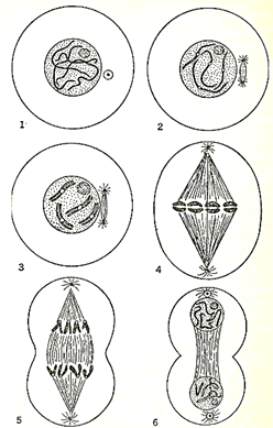
Figure 4. The stages of mitosis. (1) Chromosomes form in the nucleus in the first stage of mitosis. (2) Chromosomes begin to split into two identical halves and the aster (the small white circle outside the nucleus) spreads to opposite sides of the cell. (3) Chromosomes have doubled but remain joined at the center. (4) Chromosomes are lined up and asters have moved to opposite poles. (5) Asters pull chromosomes apart. (6) Cell begins to lengthen and ultimately will form, two new identical cells each with its own nucleus and an identical amount of chromatin as was in the mother cell in the first stage
He found that as the process of cell division began, the chromatin material coalesced into short threadlike objects which later came to be called "chromosomes" ("colored bodies"). Because these threadlike chromosomes were so characteristic a feature of cell division, Flemming named the process "mitosis" (from a Greek word for "thread").
Another change that accompanied the beginning of mitosis involved the "aster" (a Greek word meaning "star" ) . This received the name because it was a tiny dot-like object surrounded by fine threads radiating from it like the conventional rays drawn from a star. The aster divided, the two parts separating and moving to opposite sides of the cell. The fine rays passing from one aster to the other seemed to entangle the chromosomes which were grouping along the midplane of the cell.
At the crucial moment of cell division, each chromosome produced a replica of itself. The double chromosomes then pulled apart, one chromosome of each doublet going to one end of the cell and the second chromosome to the other. The cell then divided, a new membrane forming down the middle. Where there had previously been one cell, there were now two daughter cells, each with an amount of chromatin material equal (thanks to the doubling of the chromosomes) to that which had originally been present in the mother cell. Flemming published these findings in 1882.
The work was carried further by the Belgian cytologist, Eduard van Beneden (1846-1910). In 1887, he was able to demonstrate two important points about chromosomes. First, he presented evidence to show that their number was constant in the various cells of an organism, and that each species seemed to have a characteristic number. (It is now known, for instance, that each human cell contains forty-six chromosomes.)
Further, Van Beneden discovered that in the formation of the sex cells, the ova (egg cells), and spermatozoa, the division of chromosomes during one of the cell divisions was not preceded by replication. Each egg and sperm cell, therefore, received only half the usual count of chromosomes.
Once Mendel's work had been discovered by De Vries, all this work on chromosomes was suddenly illuminated. The American cytologist, Walter S. Sutton (1876-1916), pointed out in 1902 that the chromosomes behaved liked Mendel's inheritance factors. Each cell has a fixed number of pairs of chromosomes. These carry the capacity to produce physical characteristics from cell to cell, for in each cell division, the number of chromosomes is carefully conserved; each chromosome producing a replica of itself for the use of the new cell.
When an egg cell (or a sperm cell) is formed, each receives only half the usual chromosome number (one of each pair). When the fertilized ovum is formed from the union of sperm and ovum, the correct total number of chromosomes is restored. As the fertilized ovum divides and redivides to form an independently living organism, the number of chromosomes is again carefully conserved. In the new organism, however, one of each pair of chromosomes comes from the mother via the egg cell, while the second of each pair comes from the father via the sperm cell. This shuffling of chromosomes with each generation tends to bring to light those recessive characteristics earlier drowned out by a dominant characteristic. The ever-new combinations further produce over-all variations of characteristics upon which natural selection can seize.
As the twentieth century dawned, then, a sort of climax had been reached in evolution and genetics. This, however, was only to serve as a prelude to new and even more startling advances.
Date added: 2022-12-11; views: 933;