Human Ontogeny: Overview, Fertilization, and Earliest Developmental Stages
Besides gross and microscopic anatomy, the developmental history of the individual organism (ontogeny) is of key importance in understanding the human body. Ontogeny is concerned with the formation of tissues (histogenesis), organs (organogenesis), and the shape of the body (morphogenesis).
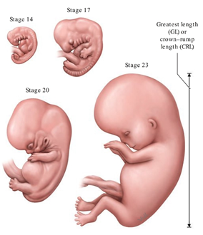
5- to 8-week-old human embryos. Streeter (1942) and O’Rahilly (1987) classified early human development and the embryonic period into 23 stages based on specimens from the Carnegie Collection. The Carnegie stages are defined by morphological characteristics that can be closely correlated with specific age (postovulatory days or weeks) and size (measured as the greatest length, excluding lower limb [GL|, or crown-rump length [CRL], see C).
Stage 14: 5th week, GL 5-7 mm, future cerebral hemispheres become identifiable.
Stage 17: 6th week, GL 11-14 mm, digital rays become visible.
Stage 20: 7th week, GL 18-22 mm, upper arms bent at the elbow, hands in a pronated position.
Stage 23: 8th week, GL 27-31 mm, eyelids fuse, external genitalia begin differentiation.
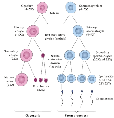
Formation of the ovum and sperm (after Sadler). During the formation of the gametes (sex cells), two successive cell divisions occur (the first and second meiotic maturation divisions). This results in cells having a chromosome set that is reduced by one half (haploid). When fertilization occurs, a diploid (full) chromosome set is restored. During meiosis, extensive chromosomal rearrangement occurs, thus recombining the internal genetic information into new and different subsets.
Oogenesis: The initial oogonia first undergo a mitotic division to form primary oocytes, which still have a diploid chromosome number (44XX). Later the primary oocytes undergo a first and second maturation division by meiosis, resulting in four haploid cells (22X): one mature ovum and three polar bodies.
Spermatogenesis: Diploid spermatogonia undergo mitosis to form primary spermatocytes (44XY). These cells then divide meiotically to form four haploid spermatids, two of which have an X chromosome (22X) and two a Y chromosome (22Y). The spermatids develop into motile spermatozoa (spermatohistogenesis).
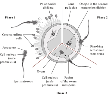
Schematic representation of the fertilization process (after Sadler). In phase 1, the spermatozoon penetrates the corona radiata cells. In phase 2, the acrosome dissolves, releasing enzymes that digest the zona pellucida. In phase 3, the cell membranes of the ovum and sperm fuse, and the spermatozoon enters the egg.
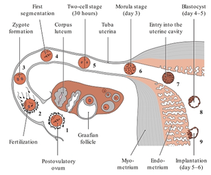
Developmental processes during the first week of development (after Sadler):
1. Ovum immediately after ovulation
2. Fertilized within approximately 12 hours
3. Male and female pronucleus with subsequent zygote formation
4. First segmentation
5. Two-cell stage
6. Morula stage
7. Entry into the uterine cavity
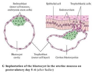
8. Blastocyst
9. Early implantation
Date added: 2023-08-28; views: 840;