The LH surge and ovulation
Once LH receptors have been fully acquired by the granulosa cells, the Graafian follicle can enter the final or preovulatory phase of growth. This terminal growth phase is signaled by a surge in gonadotrophin output, the LH surge. As the large Graafian follicle reaches maturity, estradiol output reaches a peak. In such a highly estrogenic environment, the pulse frequency of GnRH is more rapid and the sensitivity of the pituitary gonadotrope cells to GnRH is greatly enhanced. These events lead to a massive discharge of LH.
The effect of the LH surge is twofold: it causes profound changes to the structure and function of the follicle and, second, stimulates the resumption of meiosis in the oocyte.
The follicle undergoes a final rapid growth phase, mainly due to an increase in the volume of follicular fluid. Also, major changes occur in the endocrinological activity of the follicle cells.
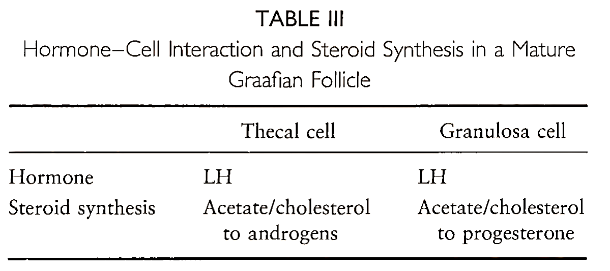
Of major importance is that the granulosa cells can no longer produce estradiol by aromatization and, thus, lose their FSH receptors. Instead, they start to synthesize progesterone through LH stimulation. These changes in hormone-cell interactions are summarized in Table III. This results in an increase in progesterone secretion from the follicle, concomitant with the rise in LH.
The production of progesterone may also be important in facilitating the positive feedback effects of estradiol on LH release. Recently, women undergoing an in vitro fertilization attempt have shown that a single injection of progesterone can elicit an LH surge. Thus, LH secretion from the pituitary is maximized, ensuring that final follicular maturation is completed.
While progesterone is being synthesized from the follicle, the chromosomes of the oocyte progress through the first meiotic division. Although the mechanisms are not fully understood, it has been proposed that rising levels of LH either inhibit the action or block the synthesis of an oocyte maturation inhibitor factor, thus allowing terminal maturation to occur.
In addition, the dose-dependent effect of LH on granulosa cell proliferation has been demonstrated in vitro, where high-dose LH caused enhanced synthesis of progesterone, suppression of aromatase activity, and inhibition of cell growth. These observations have led to the concept of an “LH ceiling hypothesis” (proposed by Steve Hillier) to explain the clinical observations demonstrating that excessive LH stimulation can disrupt normal preovulatory follicle development.
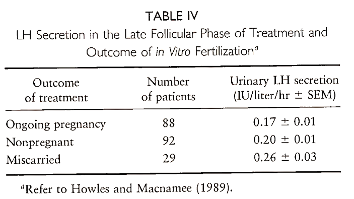
This phenomenon has been highlighted in patients undergoing superovulation for in vitro fertilization. In one study, high levels of LH were associated with failure of implantation and early pregnancy loss, whereas low levels of LH were associated with the establishment of ongoing pregnancy. Table IV summarizes the results of this study. This effect of LH on pregnancy outcome has been confirmed by other studies, including one in women not undergoing any infertility treatment. It is now widely recognized that inappropriately raised LH concentrations are a significant cause of miscarriage. Further work is required to elucidate the mechanism of LH action on oocyte maturation.
Final meiotic division of the oocyte is peculiar in that the distribution of cellular material is grossly unequal. Only a very small amount of cytoplasm accompanies one-half of the divided chromosomes; this forms what is called the first polar body, which is extruded to one side of the maturing oocyte.
Furthermore, the follicle wall undergoes dramatic changes. The rapid expansion of the follicle at this time stretches the follicle wall and, probably through the action of collagenase enzymes, particularly plasmin, and prostaglandins, the wall starts to break down. Where this occurs an outward bulge appears, the stigma, which eventually ruptures, releasing the follicle contents. By this time, the oocyte is connected to the mass of granulosa cells only by a very thin stalk of cells, which easily breaks, allowing the oocyte to be extruded in the flow of follicular fluid.
An oocyte, freshly harvested from a preovulatory follicle of a woman undergoing an in vitro fertilization treatment, is shown in Fig. 7. Note the sunburst arrangement of cumulus oophorus cells around the oocyte; this is highly characteristic of a mature human oocyte.
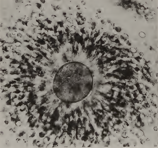
FIGURE 7. Photograph of a mature human oocyte, just after having been removed from the follicle for in vitro fertilization. The oocyte is surrounded by a mass of cumulus cells showing a dense corona radiata (sunburst appearance).
Date added: 2022-12-11; views: 1002;