Nuclear Magnetic Resonance (NMR) and Magnetic Resonance Imaging (MRI)
The phenomenon of nuclear magnetic resonance (NMR) was discovered independently in 1946 by Felix Bloch at Stanford University, and by Edward Purcell at Harvard University. For this they were jointly awarded the 1952 Nobel Prize in Physics. Chemists were quick to see the potential of NMR, as the NMR signal gives valuable information about the structure of molecules. This is because the atomic electrons, which determine the chemical properties of materials, interact with the nuclei which give rise to the NMR signals.
Much later, in 1973, Paul C. Lauterbur outlined how NMR could be used to form images for medical diagnosis. Raymond Damadian’s 1971 finding that cancerous tissue could have different NMR properties from normal tissue had prompted his work. This eventually led to a major new application of NMR in medical imaging, which we now call magnetic resonance imaging (MRI). Although Damadian and Lauterbur were both working in the U.S., much of the pioneering work needed to turn the revolutionary idea into a practical reality was done in the U.K.
in the 1970s and 1980s, at the Universities of Nottingham and Aberdeen and at the EMI Company’s London research laboratories. In 2003, the Nobel Prize in Medicine was awarded to Lauterbur, University of Illinois at Urbana, and Sir Peter Mansfield, University of Nottingham, for their discoveries, emphasizing the diagnostic importance and widespread use of NMR.
Atomic nuclei are positively charged, and some (but not all) have the quantum mechanical property termed spin. If an object is both charged and spinning, it will generate a magnetic field in the same way that a current circulating round a loop will generate a magnetic field. Thus, a nucleus that has spin can be thought of as being a tiny bar magnet. Normally, nuclear spins do not have any preferred direction of alignment.
However, if they are placed in a strong magnetic field they will tend to align with it, in much the same way as a set of compass needles align with the Earth’s magnetic field. The alignment brought about by the strong magnetic field produces an observable nuclear magnetism. Until the nuclei are placed in the strong magnetic field their magnetic dipoles are randomly oriented and their average effect is zero, so that the phenomenon of NMR cannot occur. In fact, even in a very strong magnetic field their alignment is only weak, as the nuclear magnetic moments are randomly disturbed by thermal agitation as if they were compass needles being violently shaken.
The gyroscope, or spinning top, is a good analogy for the behavior of the nuclear magnetization in a strong magnetic field. If the spinning top is perturbed from its initial alignment with the Earth’s gravitational field while supported by a table top, then it precesses around a vertical axis through its point of contact with the table. The precession is a much slower motion (in terms of revolutions per second) than the spin of the top around its own axis. Similarly, if the nuclear magnetic dipoles are perturbed from their alignment with the magnetic field, they precess around it, as shown in Figure 5.
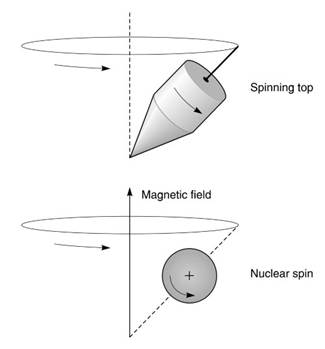
Figure 5. The spinning top is a good analogy to the behavior of the nuclear magnetism in a strong magnetic field. Once the top is pushed away from the vertical, it precesses around the gravitational field of the Earth. Similarly, once the spinning nuclei are pushed away from their alignment with the magnetic field, they also precess. The rate of precession is proportional to the strength of the magnetic field
The precession frequency f0 is often referred to as the Larmor frequency (after Joseph Larmor, an Irish physicist who investigated the behavior of electrons in magnetic fields at the end of the nineteenth century) and is given by the equation f0 = gB0. Different nuclei have different values of g. Protons have a gyro- magnetic ratio of 42.6 megahertz per tesla, while the figure for sodium is 11.3 megahertz per tesla. Thus protons (hydrogen nuclei) precess at 42.6 megahertz in a magnetic field of strength 1 tesla, while sodium nuclei (specifically of the sodium isotope with atomic weight 23) precess at only 11.3 megahertz.
It is possible to determine the frequency of a signal very accurately, by comparing it to a stable reference signal of known frequency. Thus NMR can be used to make a highly precise magnetometer—a device for measuring magnetic field strengths very accurately.
Whereas the spinning top can be pushed from the vertical by a tap of the finger, the nuclear spins have to be pushed by an oscillating magnetic field applied at right angles to the main magnetic field. The oscillation frequency has to equal the Larmor precession frequency f0, or nothing happens. This is what is meant by resonance. The field strength used in a typical NMR or MRI machine is a few tesla, so that the Larmor frequency is in the radiofrequency (RF) range. The RF magnetic field is applied by means of a tuned RF coil surrounding the patient’s body or head, with power supplied by a radio-frequency power amplifier.
The first NMR experiments in the late 1940s used the “continuous wave’’ technique, in which the frequency of the RF field is steadily increased (or decreased) while passing through resonance. In 1950 Erwin Hahn proposed the ‘‘pulsed’’ method of NMR in which the entire frequency response is obtained following a short powerful burst of transmitted RF energy called an RF magnetic field pulse.
The difference in methods can be understood by considering two possible methods of testing a church bell. In the analogy for the continuous wave method, a loudspeaker is used to produce a pure note, the frequency of which is steadily increased. When the natural tone of the bell is reached, the bell will begin to vibrate in sympathy with the applied sound. The pulsed method is faster and more direct, and can be likened to striking the bell, and listening to its note as the sound dies away.
The note contains a mixture of all the natural frequencies of the bell. Once the nuclei are precessing, their magnetic fields are also precessing, so that a tiny voltage is induced in the RF coil by the principle of electromagnetic induction. The frequency of this oscillating voltage equals the precession frequency. If a number of different frequencies are present in the signal, because the nuclei are in a number of different electronic environments, then the different frequency components have to be separated by a mathematical process called Fourier transformation, carried out by a computer.
The use of NMR to investigate the atomic and molecular structure of materials is called NMR spectroscopy. During the 1950s and 1960s, NMR spectroscopy became a widely used technique for the nondestructive analysis of small samples, particularly of liquids. The electron cloud surrounding the nuclei within individual molecules modifies the strength of the magnetic fields sensed by the NMR sensitive nuclei, and hence changes the frequency of the NMR signal that the nuclei emit. In addition, neighboring nuclei can also influence each other.
Pulses of RF energy are used to perturb the NMR sensitive nuclei, and sensitive RF receivers are used to pick up the signals they give out. The pulsed method allied with computerized Fourier transformation revolutionized NMR spectroscopy in the 1970s. The ‘‘spectrum’’ is a plot of signal strength versus NMR frequency and contains much useful information about the chemical structure of the material under testing. NMR spectroscopy is now widely used in the fields of biomaterials, polymer chemistry, and solid-state physics.
Pulsed NMR techniques can also be used to form medical images. MRI normally uses signals arising from hydrogen nuclei, because they are so much more abundant in the body than any other NMR-sensitive nucleus and therefore give a measurable signal even from small volumes of tissue. However, phosphorus and sodium MRI is also possible. Hydrogen nuclei that are MRI visible occur predominantly in water in the tissues and in body fat.
Figure 6 shows a patient lying on a table, about to be moved into an MRI scanner, with the head resting inside the RF head coil. The electromagnet has a large tunnel in which the patient lies and uses a special wire immersed in liquid helium so that the wire superconducts. Thus the magnet does not need any electrical power to generate the field. To form an image, it is essential to have a method of determining the position of the nuclear spins within the magnet.
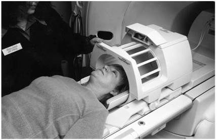
Figure 6. A patient lies on a table before being transferred into the MRI scanner. Her head is being imaged and has to be placed inside the radio-frequency head coil beforehand
This is accomplished by field gradients, a method proposed by Paul Lauterbur of the State University of New York at Stony Brook in 1973. A field gradient coil modifies the strength of the main magnetic field along a particular direction so that it varies in a linear way. Three independent field gradient coils are used in order to generate the gradients in x, y, or z directions (along the magnet tunnel, left to right across the tunnel, and vertically). When a field gradient is on, therefore, the Larmor frequency will also vary in a linear fashion along the direction of the field gradient.
The first step in imaging is to tip the nuclear spins away from their alignment along the main magnetic field using a short-pulsed RF magnetic field oscillating at the Larmor frequency, as explained above. If the pulse is applied in the presence of a field gradient along the patient (along z), then only one transverse plane across the body will respond, as only one plane has a Larmor frequency that matches the RF frequency.
The imaging process is now simplified to imaging a two-dimensional slice in an x-y plane, rather than imaging the entire three-dimensional body. The gradient along the patient is switched off, and a gradient switched on across the body (along x). This makes the nuclei precess faster on one side of the body, and more slowly on the other. Thus the frequency of the NMR signals varies in a linear way across the body, so that a particular NMR signal frequency corresponds to a particular left to right (x) position. In order to form the image, the vertical (y) position also has to be encoded onto the NMR signal.
Although a full discussion is impossible here, the technique involves repeating the above process many times, with a small pulse applied to the vertical y gradient just before the signal is collected and with the amplitude of the pulse being stepped up between each repetition. This is the ‘‘spin-warp’’ imaging method, invented by William Edelstein and James Hutchison of Aberdeen University in 1980. It is still the most widely used imaging method today.
Modern MRI scanners rely on fast computers to carry out the large number of digital Fourier transforms necessary to form the images and to control very precisely the timing of the pulsed currents flowing in the gradient and RF coils.
Figure 7 shows a midline slice through the center of a human head on an MRI image. Note the excellent anatomical detail achieved. Areas with no protons, such as the air-filled sinuses behind the nose, give no signal and appear dark. Dense hard bone in the skull also has few protons and gives no signal. Watery fluid such as the cerebrospinal fluid around the spinal cord gives a different signal (brighter in this case) from that of the soft tissue of the brain because the NMR properties of the water protons are affected by their biophysical environment.
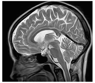
Figure 7. A section of the brain imaged by MRI. Note the excellent anatomical detail the scan gives
In other words, image detail arises from tissue structure on a microscopic level, as well as from differences in proton density. This is the particular strength of MRI in medical diagnosis because many disease processes, including cancer can therefore be visualized.
A recent development in MRI is its use to repeatedly image the brain every 2 or 3 seconds as it performs different tasks or responds to various stimuli, a technique called functional MRI (fMRI), developed independently by Seiji Ogawa of AT&T Bell Laboratories in New Jersey and John Belliveau at Massachusetts General Hospital in Boston in the early 1990s. This requires a highperformance scanner capable of performing the very high-speed imaging technique of ‘‘echoplanar’’ imaging, invented by Peter Mansfield at Nottingham University in 1977.
The fMRI technique relies on the oxygen-dependent magnetic effect of the iron atoms contained in the hemoglobin molecules of the blood. When part of the brain is active, its oxygen consumption increases, thus stimulating a large increase in the local blood supply, and the oxygen concentration in the tiny blood-filled capillary vessels is raised. For the particular rapid-imaging method used in fMRI, this causes a small increase in the signal detected.
In its simplest form, fMRI alternates periods of rest, lasting 30 seconds or so, with equal periods of activation. Relatively large signal changes of a few percent can be obtained with visual activation using flashing lights or rapidly alternating checkerboard patterns (black squares turning to white and vice-versa), while more subtle cognitive tasks involving memory or reasoning give smaller changes. In other words, MRI can image your thoughts, an application never dreamt of by Felix Bloch and Edward Purcell in 1946.
Date added: 2023-10-26; views: 707;