Drawing of the structural components of the basement membrane of epithelium
Fig. 2-21. Drawing of the structural components of the basement membrane of epithelium. Each cell has an anionic charge on its surface from the presence of charged groups of carbohydrates in the glycocalyx (cell coat). The basement membrane consists of three components. The lamina rara, an electron-lucent zone rich in polysaccharides, is located between the cell membrane and the second component, the lamina densa (basal lamina). The lamina densa consists of a lattice of type IV collagen and laminin to which are bound sulfated proteoglycans and other minor proteins. The third component, the reticular lamina, consists of type III collagen fibrils and anchoring fibrils that strengthen the interface between the lamina densa and underlying reticular lamina.

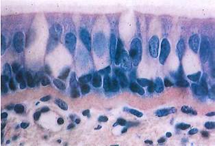
Fig. 2-22. Pseudostratified columnar epithelium with cilia and goblet cells seen by LM. The basement membrane is thick, smooth, and pink and lies immediately below the epithelium. (H&E; X450.).

Fig. 2-23. ТЕМ of the basal region of a stratified squamous epithelium showing the basement membrane and underlying connective tissue. Immediately below the plasmalemma of the epithelial cell, the lamina rara and lamina densa (basal lamina) of the basement membrane are clearly seen. Anchoring fibrils extend from the lamina densa into the adjacent connective tissue, where they intercalate with reticular fibers. (X 13,000.).
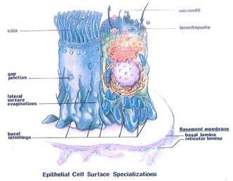
Fig. 2-24. Surface specializations characteristically found in epithelial cells include luminal surface projections such as cilia or microvilli, junctions to regulate permeability (zonula occludens), adhesion (desmosome), communication (gap), amplification of basal membrane related to ion transport (parietal cell in stomach and proximal or distal tubule cells in kidney), and modifications of the basal surface to facilitate attachment to the basement membrane.
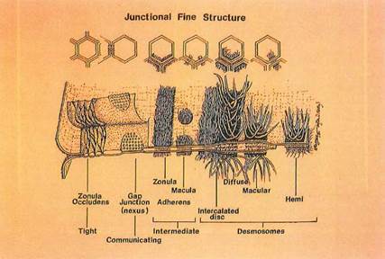
Fig. 2-25. Fine structure of different types of junctions. The zonula occludens junction regulates permeability between adjacent epithelial cells in most epithelia, except for stratified squamous where permeability is regulated by an intercellular lipid barrier. Gap junctions facilitate the intercellular passage of small molecules and are found in excitable cells (e.g,, nerve and cardiac or smooth muscle cells) as well as nonexcitable cells (e.g., osteocytes, Sertoli cells, granulosa cells, intestinal epithelial cells). A variety of junctions using either actin (zonula adherens) or intermediate filaments have been described in different cell types.
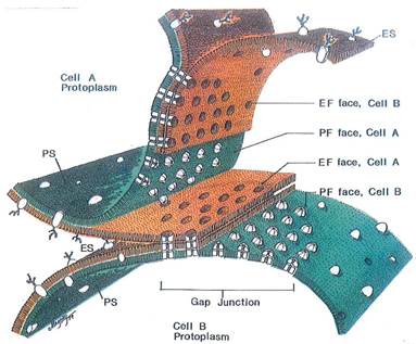
Fig. 2-26. Freeze fracture passing from the plasmalemma of cell В to that of cell A. Note that the fracture plane occurs within the lipid bilayer of each cell membrane. Intramembranous particles are usually more numerous in the protoplasmic fracture membrane face (PF face), where they are often seen as groupings of hexagonally packed intramembranous particles forming gap junctions. Gap junctions control transport of ions and small molecules between adjacent coupled cells, resulting in the communication of electrical and metabolic signals. Such ionic or molecular transport via gap junctions is thought to occur through the central channel formed by opposing connexons in each cell membrane.
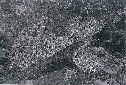
Fig. 2-27. FF of a gap junction between tumor cells. Both the protoplasmic (P) and extracellular (E) faces are seen, and gap junctions appear in both, (x 25,000.).
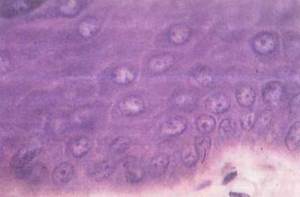
Fig. 2-28. Stratified squamous epithelium of shin. By LM the cells of this layer appear to have spiny borders, which, by ТЕМ, are seen to be points of intercellular adhesion called desmosomes. However, the impression gained by LM resulted in the name of "stratum spinosum” for this layer of skin. (H&E; X 900.).
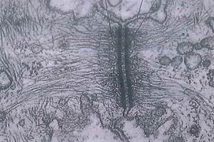
Fig. 2-29. Desmosomes, as viewed by ТЕМ, exhibiting tonofilaments embedded in electron-dense plaque at the junction and extending into the cytoplasm. Tonofilaments do not cross intercellular space, although recent studies suggest that some particles called membrane linkers may somehow be attached to intramembranous particles (see Fig. 2-30). (X 56,700.).
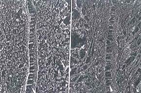
Fig. 2-30. Deep etch ТЕМ of desmosomes revealing tonofilaments in a cross fracture of cytoplasm. Dense masses of filaments can be seen near the plasmalemma, as can the presence of membrane linkers (arrowheads) crossing the extracellular space between the adjacent cells. (x 90,000.).

Fig. 2-31. Terminal bars, or junctional complexes that prevent luminal contents from penetrating lateral intercellular space. They are seen here by LM as darkly stained spots (arrowhead) at the lateral cell membranes immediately below the microvillous border. (H&E; X 1,300.).
Date added: 2022-12-11; views: 1109;