ТЕМ of the simple columnar epithelium in the small intestine. ТЕМ of transitional epithelium from the bladder
Fig. 2-7. ТЕМ of the simple columnar epithelium in the small intestine. Note the goblet cell in the center of the micrograph and numerous intraepithelial lymphocytes between it and adjacent absorptive cells. A fenestrated capillary can be seen in the lamina propria beneath the goblet cell. (x 4,000.).

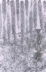
Fig. 2-8. Higher magnification ТЕМ of the microvillous border of simple columnar epithelial cells in the small intestine. Bundles of actin filaments in the core of each microvillus (arrowhead) insert into the terminal web region, as do tonofilaments (intermediate filaments) from the desmosome (lower right) and actin filaments from the zonula adherens (asterisk) of the junctional complex. (X 60,000.).
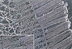
Fig. 2-9. ТЕМ of a deep etch preparation of the terminal web region of a cell and its microvillous border. Note (as in Fig. 2-8) the relationships between actin in the core of each microvillus and the network of intermediate filaments in the terminal web (T). Inset (lower left) shows that tightly packed bundles of actin filaments in each microvillus are enmeshed by a fine network of cross-linked filaments. (X 38,800.).
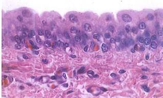
Fig. 2-12. Transitional epithelium of the urinary bladder seen by LM. Umbrella cells, at the lumen, will stretch and flatten as the bladder fills. Red blood cells within capillaries are separated from the epithelium by the basement membranes of both the epithelium and the capillary endothelium. (H&E; X500.).
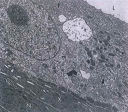
Fig. 2-13. ТЕМ of transitional epithelium from the bladder. Umbrella cells form the luminal surface of the epithelium but do not contact the basement membrane. These cells contain discoidal vesicles (arrow) associated with endocytosis/exocytosis of luminal membrane when the cell undergoes shape change (squamous-cuboidal-squamous, etc.) because of stretch or relaxation of the bladder. (X 3,100.).
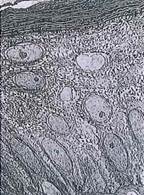
Fig. 2-15. ТЕМ of stratified squamous keratinized epithelium from thin skin. The basement membrane is seen at the bottom right. Note the change in cell shape as the cells migrate from the basement membrane toward the surface. Desmosomes can be seen as dense spots at the cell margins. (x 1,500.).
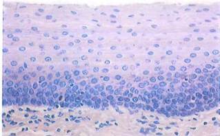
Fig. 2-16. LM of stratified squamous nonkeratinized epithelium of the esophagus. This thick epithelium contains many layers of cells. The long axes of the surface cells are parallel to the basement membrane, while those of basal cells are perpendicular to it. The nuclei of cells of the upper layers are ovoid, not flattened, and the luminal surface lacks keratinized squames. (H&E; X 350.).
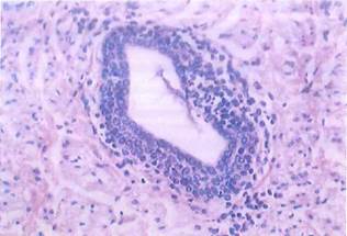
Fig. 2-17. LM of a duct lined by stratified cuboidal epithelium. Small lymphocytes are present in the epithelium and the adjacent connective tissue. (H&E; X250.).
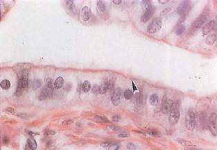
Fig. 2-18. LM of simple columnar ciliated epithelium from the uterine tube. The basal bodies of the cilia form an acidophilic band in the apical cytoplasm (arrowhead). At this magnification, the cilia look like thin filaments extending into the lumen. (H&E; X600.).
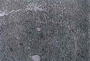
Fig. 2-19. ТЕМ of simple columnar epithelium in the uterine tube showing the apical surface of ciliated cells bordering the lumen. The cilia arise from basal bodies located immediately beneath the cell surface. (X2,100.).
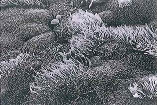
Fig. 2-20. SEM of cilia and microvilli on the luminal surface of rat trachea. Note that the apical surfaces of cells with microvilli or cilia may be distinguished from one another because of the greater length of cilia (about 8 to 10 |im) versus that of microvilli (about 1 (xm). (x 1,450.).
Date added: 2022-12-11; views: 1169;