Composite Cell. Three-dimensional representation of simple columnar cells showing organelles
Cells Compose the Four Basic Tissues of Body: epithelium, connective tissue, muscle, and nerve. Common to all mature cells, or cells at some time in their development, are nuclei and cytoplasm containing membranous compartments and cytoskeletal components. Cells vary considerably in size, shape, type, and distribution of organelles and, consequently, function. Despite these differences, by understanding the role of each organelle one can deduce general structural/functional correlates that lead to a clearer understanding of how cells work.
To obtain a better concept of the function of cells and their organelles, it is helpful to examine cells by a variety of methods. Light microscopy (LM) can be used with routine histologic stains (hematoxylin and eosin; H&E) to obtain information on the biochemical function and distribution of cellular components. The basophilic staining of nuclei by hematoxylin provides information on the distribution of inactive heterochromatin and the number of nucleoli, while cytoplasmic basophilia may reflect the presence of free or bound ribosomes (rough endoplasmic reticulum, RER) associated with protein synthesis or, in some other instances, the synthesis of sulfated mucosubstance destined for secretion.
The intensity of cytoplasmic staining with eosin (acidophilia) generally reflects the relative abundance of cationic proteins. Mitochondria contain cationic proteins, such as cytochromes, which leads to intense cytoplasmic eosin- ophilia in cells involved in ion transport since they use abundant mitochondria to produce energy (ATP) to drive membrane pumps. Transmission electron microscopy (ТЕМ) permits examination of the ultrastructure of organelles and cells, but interpretation of the fine structure, particularly in three dimensions, is hampered by the use of ultrathin sections needed for penetration of the sample by the electron beam.
Visualization of the three-dimensional appearance of the surfaces of organelles, cells, and tissues can be better accomplished by using scanning electron microscopy (SEM); however, this method by itself does not necessarily provide information on the internal fine structure of cells. The freeze-fracture (FF) technique supplies information on the structure of membranes, particularly the distribution of intramembranous proteins related to the formation of cell junctions, but yields little information on the general structure of cells. Despite the limitations of each of the methods just described, when used collectively they enable us to obtain a comprehensive view of the structure and function of organelles and cells.
A typical cell will include the nucleus, free ribosomes, polyribosomes, and a variety of membranous organelles including rough endoplasmic reticulum (RER), smooth endoplasmic reticulum (SER), Golgi apparatus, mitochondria, peroxisomes, and components of the lysosomal system. Cytoskeletal organelles present within the cell include free microtubules and microtubular arrays including centrioles, basal bodies, flagella, and cilia; contractile proteins such as actin and myosin; and structural proteins comprising the intermediate filaments (desmin, glial fibrillary acidic protein, keratin, neurofilaments, nuclear lamins, and vimentin). Nonorganelle components that may be present within the cytoplasm of cells are glycogen, lipid inclusions, and pigment.
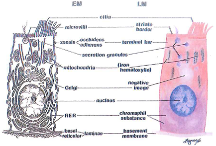
Composite Cell. Fig. 1-1. Drawing of a composite, polarized epithelial cell comparing ultrastructural components as viewed by transmission electron microscopy (ТЕМ) and light microscopy (LM). Mitochondria can be visualized at the LM level by the use of special stains, in this case, iron hematoxylin.
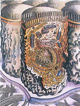
Fig. 1-2. Three-dimensional representation of simple columnar cells showing organelles, microvillous border (also known as brush or striate border), and basement membrane. The lateral borders of cells interdigitate, and junctional complexes at the apical borders connect them into a tissue called epithelium, which serves as a permeability barrier between body fluids and the external environment. Beneath the basement membrane is a connective tissue compartment containing collagen fibers and amorphous ground substance.
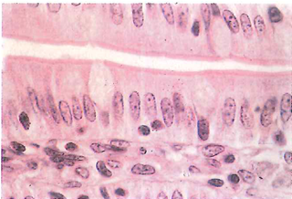
Fig. 1-3. Intestinal epithelium of simple columnar cells as seen by LM. Visible in the epithelium are mucus-secreting goblet cells, lymphocytes between the lateral borders of columnar cells, a microvillous border adjacent to the lumen, and the basement membrane subtended by connective tissue called the lamina propria. (H&E; X 1,600.)
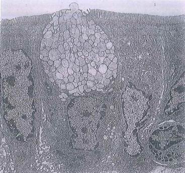
Fig. 1-4. Intestinal epithelium with a goblet cell seen by transmission electron micrograph (ТЕМ). Compare with Figs. 1-2 and 1-3. Identify in the epithelium the mucus cell containing secretion granules, a microvillous border, nuclei, mitochondria, lateral cell borders, Golgi regions, and basement membrane. A wandering granulocyte (white blood cell) is seen between the epithelial cells at lower right, (x 4,000.).
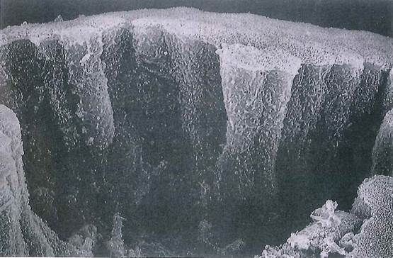
Fig. 1-5. Simple columnar epithelial cells of the gallbladder seen by scanning electron micrograph (SEM). Note that evaginations of lateral cell surfaces interdigitate with the membranes of adjacent cells. See Fig. 1-4 for the appearance of these membrane projections by ТЕМ. (X 2,400.).
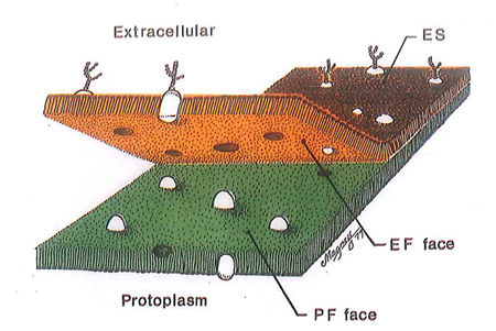
Fig. 1-6. Drawing of a cell membrane (plasmalemma) that has been freeze-fractured (FF). Note that the fracture plane passes through the lipid bilayer and that intramembranous particles tend to remain with the protoplasmic face (PF face). The extracellular face (EF face) is generally characterized by pits or depressions that remain when intramembranous particles pull free as a result of the fracture. ES, Extracellular surface.
Date added: 2022-12-11; views: 2353;