Classification of the eight subdivisions of epithelia
Epithelium covers the free surfaces of the body, lines body tubes and cavities, forms glands, and assumes many different arrangements to meet a variety of functions (i.e., protection, absorption, secretion, reproduction, excretion, digestion, lubrication, and sensory reception). The diversity of the epithelia associated with these functions can be linked to their use of surface modifications (of epithelial cells) and adaptation of cellular organelles to meet the unique functional requirement.
Classification of epithelia is based on the numbers of layers of cells (simple, one layer; stratified, two or more layers), the shape of the luminal or surface layer of cells (squamous, cuboidal, columnar), and the presence of surface specializations, such as cilia or keratin. Clusters of epithelial cells designed for secretion and connected to epithelial surfaces by ducts formed of epithelial cells are called exocrine glands. Masses of epithelial cells, or in some cases even individual cells, that in the absence of ducts release their secretory product into the vascular system are called endocrine glands.
Characteristics of epithelia include the presence of highly polarized contiguous cells, the absence of intercellular substance and blood vessels, and surface specializations of their intercellular, basal, and luminal surfaces. Depending on the function of the epithelium, epithelial cells may be attached to one another by specialized intercellular junctions associated with cellular adhesion (stratified epithelia), permeability (simple epithelia), cellular communication (glandular epithelia), or, in many instances, a combination of these. The basement membrane separates the basal surface of the epithelium from the underlying connective tissue and is composed of two elements, the basal lamina (type IV collagen) and a reticular lamina of variable thickness containing reticular fibers (type III collagen).
The basal lamina of the epithelium serves both as a site of physical attachment for the epithelium and, in some instances, as a specialized permeable interface between the body and its environment (i.e,, kidney and lung). The reticular lamina is bound to, and merges with, the underlying connective tissue, which serves as both a structural and a nutritional support for the avascular epithelium. Epithelial cells involved in ion transport may possess dramatic infoldings of the basal surface, which increase the surface area available for the movement of ions by membrane pumps.
Epithelial cells may possess two types of membrane specializations on the luminal surface: microvilli and cilia. Microvilli are finger-like projections of the luminal surface that amplify the surface area of the cell. Cells involved in membrane transport (i.e., small molecules such as sugars and amino acids) may have numerous densely packed microvilli, referred to by classical histologists as a striate or brush border, depending on their location in small intestine or kidney, respectively. Each microvillus contains a core of actin filaments whose state of polymerization may regulate its length and thereby the total area of the luminal surface.
Microvilli are usually 0.1 pm in diameter and 0.5 to 1.0 pm in length. In the male reproductive tract, they are seen as exceedingly long projections called stereocilia, measuring up to 8 to 10 pm in length. Cilia are motile luminal specializations associated with the transport of extracellular materials such as mucus, debris, or cells. Each cilium is composed of nine double microtubules and a pair of central microtubules. Cilia are approximately 8 pm long and 0.2 pm wide and may be numerous on cellular surfaces, whereas a flagellum usually exceeds 30 or 40 pm in length and occurs only in sperm cells.

Fig. 2-1. Classification of the eight subdivisions of epithelia. Epithelium is a tissue that covers the free surfaces of the body. The cells are contiguous and rest on a basement membrane, which acts as an interface with the underlying connective tissue. Epithelia are classified as simple (containing one layer of cells), or stratified (multiple layers are present). The specific name of the epithelium reflects the shape of the cells lining the luminal surface (squamous, cuboidal, columnar, or transitional). Pseudostratified epithelium refers to the apparent presence of multiple cell layers; however, all cells are attached to the basement membrane, with only tall columnar cells reaching and forming the luminal surface. Transitional epithelia found in the urinary system are named for the changes in shape of surface cells during relaxed-stretched states related to storage of urine.

Fig. 2-2. LM showing a simple squamous epithelium lining Bowman's space of the renal corpuscle and a simple cuboidal epithelium forming the distal convoluted tubule in the kidney. (H&E; X 500.).
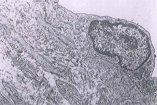
Fig. 2-3. A simple squamous cell seen by ТЕМ. This cell is part of the mesothelium forming the visceral peritoneum of the intestine. Elements of connective tissue and smooth muscle are present beneath the basement membrane, (x 4,600.).
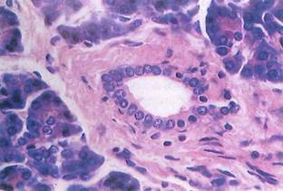
Fig. 2-4. Simple cuboidal epithelium of a pancreatic duct seen by LM. The duct is surrounded by connective tissue and pancreatic acini. (H&E; X350.).

Fig. 2-5. Cross, section of a pancreatic duct by ТЕМ demonstrating simple cuboidal epithelium. Individual cells are approximately equal in height and width. (X 3,400.).
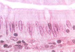
Fig. 2-6. LM of the small intestine revealing a simple columnar epithelium with goblet cells. Mucous granules in the goblet cells are unstained owing to the method used to fix the tissue. Intraepithelial lymphocytes are also present. (H&E; X 1,400.).
Date added: 2022-12-11; views: 1178;