Cartilage is a specialized form of connective tissue designed for tissue support and weight bearing
Cartilage is a specialized form of connective tissue designed for tissue support and weight bearing. Like other connective tissues, it consists of cells and extracellular matrix. Unlike other connective tissues, the extracellular matrix is modified to form a semirigid gel. Cells embedded within the matrix occupy spaces called lacunae and are called chondrocytes, whereas peripheral, formative cells are called chondroblasts.
The extensive extracellular matrix is produced by chondrocytes/chondroblasts and consists of connective tissue fibers, proteoglycans, and chondronectin. Depending on the type of cartilage, connective tissue fibers in the matrix may include type II collagen fibrils exhibiting 64 nm periodicity but not aggregating to form bundles visible by LM, elastic fibers, or type I collagen. Nonfibrous components of the extracellular matrix include sulfated proteoglycans and hyaluronic acid, which together form proteoglycan aggregates and chondronectin. The abundant proteoglycans in cartilage are responsible for the high water content and, together with collagen for reinforcement, give cartilage its solid, yet resilient properties.
Chondronectin, a structural glycoprotein, enhances the adherence of chondrocytes to the extracellular matrix. Production of extracellular matrix results in a peripheral zone surrounding each chondrocyte that is rich in proteoglycans and deficient in collagen. This zone is called the capsular or territorial matrix because of its intense basophilic staining with hematoxylin and eosin. Located between adjacent chondrocytes is the interterritorial matrix, which exhibits less basophilic staining because of its lower content of proteoglycans.
Variations in the cellular and fibrous components of the extracellular matrix result in the classification of three different types of cartilage. The most common type, hyaline cartilage, contains type II collagen and a high proportion of sulfated proteoglycans that, because of their similar refractive index, make the collagen fibrilsundetectable by LM. Hyaline cartilage serves as a temporary skeleton in the embryo and forms the epiphyseal plate during the growth of long bones. It is also present in the supporting cartilages of the respiratory tract, the costal cartilages of ribs, and the articular surfaces of bones.
Elastic cartilage contains an abundance of elastic fibers within the matrix in addition to type II collagen. Elastic cartilage is more pliable and deformable than hyaline cartilage. It is located in the external ear, auditory tube, epiglottis, and larynx. Both hyaline and elastic cartilage arise from mesenchymal precursors and are therefore surrounded by an interface of dense connective tissue called a perichondrium. Cartilage is a unique connective tissue; it is avascular because of its production of factors inhibiting angiogenesis, and therefore it must derive its nutritional support by diffusion from blood vessels located in the perichondrium.
Fibrocartilage, unlike hyaline or elastic cartilage, does not differentiate from mesenchymal cells but, instead, differentiates in areas of dense connective tissue subjected to stress of the demands of weight bearing. Thus fibrocartilage lacks a perichondrium, and the extracellular matrix of fibrocartilage contains a dense network of coarse, eosinophilic, type I collagen fibers easily visible by LM. Both the cell density and the amount of proteoglycans in fibrocartilage are less than that of either hyaline or elastic cartilage. Fibrocartilage can be found in intervertebral disks, pubic symphysis, menisci and ligaments of joints, and at the insertion of tendons or ligaments into bone.
Cartilage undergoes growth by two different mechanisms: appositional growth, a result of the differentiation of osteoprogenitor cells in the perichondrium into chondroblasts, and interstitial growth, the mitotic division of chondrocytes. Clusters of cells derived from a single chondrocyte within matrix may be designated as isogenous cell nests or groups. In cartilage of the epiphyseal plate cell the cells are arranged in rows.
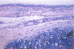
Fig. 4-1. A portion of hyaline cartilage (c) is seen beneath the tracheal epithelium in this LM. An eosinophilic perichondrium surrounds the matrix of the cartilage, which appears basophilic. (H&E; X300.).

Fig. 4-2. Compare this SEM of tracheal cartilage to the previous LM of the same tissue (Fig. 4-1). Identify the perichondrium, the cartilage matrix, and cavities within the matrix called lacunae that house chondrocytes. (x400.).
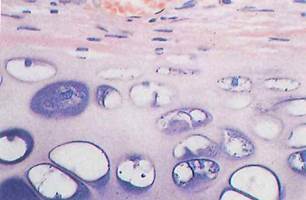
Fig. 4-3. Chondrogenic cells and chondroblasts are located in the basal portion of the perichondrium of hyaline cartilage. (H&E; X 750.).
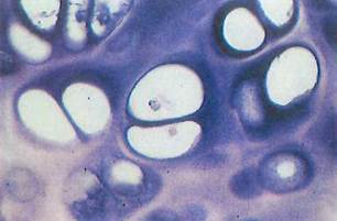
Fig. 4-4. In hyaline cartilage, chondrocytes or groups of chondrocytes called isogenous cell nests are housed in lacunae that are surrounded by basophilic territorial matrix. The chondrocytes have shrunk away from the lacuna walls as a result of the process of fixation. (H&E; X 1,200.).
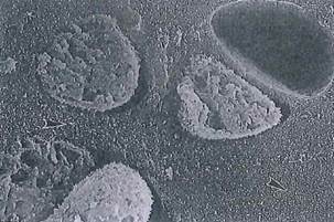
Fig. 4-5. SEM of isogenous cell nests shows chondrocytes covered by short microvilli occupying the entire lacuna. The interterritorial matrix contains thin collagen fibrils (arrowheads). (x 3,000.).
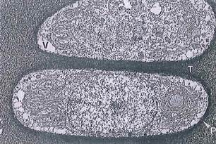
Fig. 4-6. This electron micrograph (ТЕМ) of hyaline cartilage reveals proliferating chondrocytes that were fixed in the presence of ruthenium hexamine trichloride to stabilize proteoglycans in the matrix. P, Pericellular (capsular) matrix; T, interterritorial matrix; V, artifactual vacuoles created by fixation. (x 3,900.).
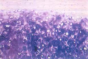
Fig. 4-7. This LM shows elastic cartilage from the epiglottis. Elastic fibers look unstained (or light pink in older specimens) and weave between lacunae. The basophilia of the matrix is due to sulfated glycosaminoglycans. Perichondrium is at the top of the section. (H&E; X300.).

Fig. 4-8. In this LM of fibrocartilage observe the shape of the cells, their relative numbers, and the presence of fiber bundles. (H&E; X 300.).
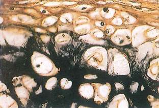
Fig. 4-9. In this high-magnification LM of elastic cartilage and perichondrium the elastic fibers are selectively stained black. Compare it to Fig. 4-7. (Verhoeff stain; x 750.).

Fig. 4-10. A high-magnification LM of fibrocartilage demonstrates cell nests and their territorial matrix. (Compare to Fig. 4-8.) (H&E; x 750.).
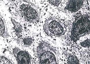
Fig. 4-11. ТЕМ of chondrocytes in elastic cartilage. The interterritorial matrix contains slender collagen fibrils (arrowhead), proteoglycan particles, and strongly electron-dense elastic fibers (ef), some of which are associated with the surfaces of chrondrocytes. (x 3,200).

Fig. 4-12. By ТЕМ note the presence of aggregations of collagen fibers in the matrix of fibrocartilage. Compare this image with the ТЕМ of hyaline cartilage (Fig. 4-6) showing dispersed collagen fibrils. ER, Endoplasmic reticulum; G, Golgi; Mv, matrix vesicles. (X 5,600.).
Date added: 2022-12-11; views: 1040;