LM of loose connective tissue. LM of adipose connective tissue
Fig. 3-12. LM of loose connective tissue. Cells and collagen fibers are equally abundant in loose connective tissue. It serves as the interface or packing substance between all other tissues or organs in the body. Compare this to other types of connective tissue on this page. Contrast the relative abundance of cells and fibers and their arrangement. (H&E; X 100.).
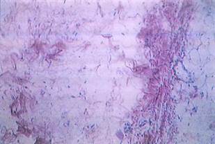
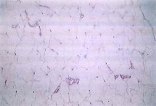
Fig. 3-13. LM of adipose connective tissue. The predominant cell is the adipocyte, or fat cell, surrounded by a delicate network of reticular fibers that attaches it to neighboring cells or tissues. Large bundles of collagen are absent. (H&E; x 100.).
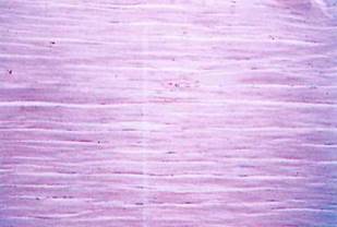
Fig. 3-14. LM of tendon, which is dense regular connective tissue. Large bundles of collagen (type I) fibers are oriented in the same plane. Cellular elements are few and consist mainly of fibroblasts. (H&E; x 100.).
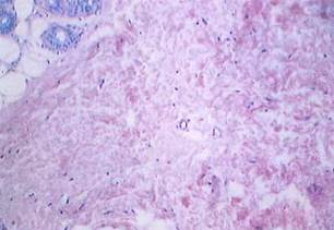
Fig. 3-15. LM showing the dermis of skin, which is dense irregular connective tissue. Collagen bundles predominate but are oriented in several different planes, giving the appearance of an irregular arrangement. (H&E; x 100.).
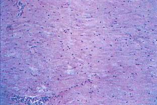
Fig. 3-16. LM of the ligamentum nuchae (interspinous ligament) showing that it consists of a dense regular arrangement of connective tissue fibers with compact masses of elastic fibers predominating. The presence of elastic fibers is masked by the eosinophilic staining of collagen. (H&E; x 100.).
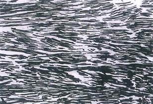
Fig. 3-17. LM of the ligamentum nuchae with an elastin stain, which reveals the presence of large masses of elastic fibers oriented in the same plane. Compare this to Fig. 3-16. (Verhoeff stain; x 100.).
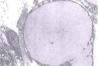
Fig. 3-20. ТЕМ of an adipocyte from the subcutaneous tissue of a 6-month-old fetus. The nucleus (N) is pressed to one side of the cell by the large lipid droplet (Ld). The cytoplasm is reduced to a thin rim containing small Golgi complexes and mitochondria. Collagen fibers (Cf) and fibroblasts (Fb) are seen at left. (X 1,600.).
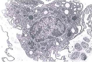
Fig. 3-21. A free macrophage (ТЕМ) exhibiting numerous cytoplasmic infoldings, vacuoles, coated vesicles, and residual bodies. Examples of collagen fibers and unmyelinated nerve fibers are also seen in the extracellular matrix. (x 6,000.).
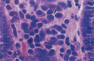
Fig. 3-26. Plasma cells with eccentric nuclei (arrowhead) within connective tissue. These cells contain nuclei with a "clock-face" configuration of condensed chromatin and basophilic cytoplasm. By LM, plasma cells are often seen beneath mucosal surfaces (as here, in the intestine) and in lymphoid tissue. (H&E; X 500.).
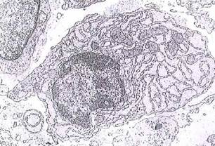
Fig. 3-27. A plasma cell by ТЕМ exhibiting dilated cisternae of rough endoplasmic reticulum. This cell is active in secretion of protein (antibody). Nuclear patterns of euchromatin and heterochromatin are evident, (x 3,200.).
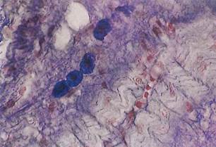
Fig. 3-28. LM of four mast cells in connective tissue. Note large, densely packed granules within the cytoplasm and the central nucleus. (Aldehyde fuchsin stain; x 500.).
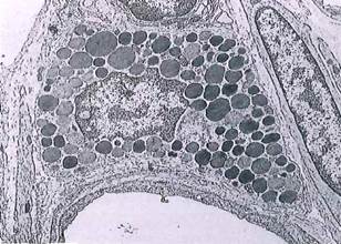
Fig. 3-29. ТЕМ of a mast cell. Granules contain histamine, which increases the permeability of small venules producing edema, and heparin, an anticoagulant. A portion of a capillary is seen at upper right. (X 3,200.).
Date added: 2022-12-11; views: 1085;