Diagram illustrating the composition of ground substance in the extracellular matrix
Fig. 3-3. Diagram illustrating the composition of ground substance in the extracellular matrix. Considerable heterogeneity in proteoglycan size and structure exists between different tissues owing to variations in the number, size, or degree of sulfation of GAG chains. The proteoglycan aggregate illustrated in this frame is from cartilage. Recently fibronectin, chondronectin, and laminin (all glycoproteins) have been identified as structural elements of the connective tissue matrix. Fibronectin is a major surface glycoprotein of the fibroblast, but it may be produced by other mesenchymally derived cells, by epithelial and endothelial cells, and by some marrowcell types. It is also found in plasma and is sometimes called cold-insoluble globulin. Laminin is a constituent of basement membranes; chondronectin is found within the matrix of cartilage.
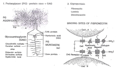
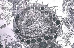
Fig. 3-2. ТЕМ of a mast cell in connective tissue. Note the presence and size of proteoglycans (drawn in ink) in the extracellular space. Examples of collagen fibrils and fibers can also be seen in the extracellular space. (X 10,000.).
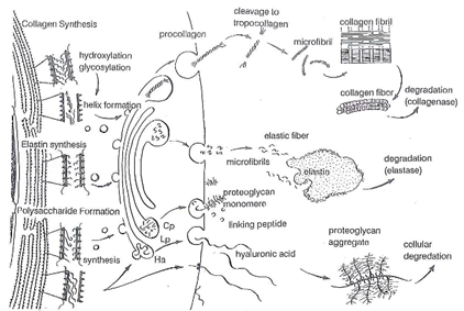
Fig. 3-3. Drawing of a fibroblast illustrating steps in the intracellular synthesis and extracellular formation of collagen, elastic .fibers, and proteoglycans. The intracellular formation of both collagen and elastin results in the secretion of a pro form of the molecule. Procollagen or proelastin are modified extracellularly to permit formation of fibers. Exopeptidases clip off the amino- or carboxyl- termini of procollagen, thereby making the molecule more insoluble, Cross-linking of adjacent tropocollagen molecules is facilitated by lysyl oxidase, and in the case of elastin, lysine residues are cross-linked to form the unusual amino acids, desmosine and isodesmosine. Glycosaminoglycans are secreted by vectorial transport through the Golgi apparatus, whereas hyaluronic acid may follow a similar pathway or possibly may be released via a transporter molecule as occurs in microorganisms. Small amounts of proteoglycans and hyaluronic acid may also be associated with the extracellular surface of cell membranes.
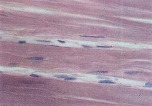
Fig. 3-4. LM of tendon in longitudinal section demonstrating a dense array of collagen fibers and interspersed fibroblasts. (H&E; x 500.)
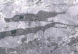
Fig. 3-5. ТЕМ of fibroblasts and collagen. Note the size and plane of section of the collagen fibers, (x 3,600.).
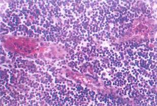
Fig. 3-6. LM of a section of lymph node stained with H&E. The lymph node is an organ composed mainly of lymphocytes, which make up the parenchyma or functional part of the organ, suspended in a net or framework of reticular fibers called the stoma. Reticular fibers are unstained in this section and therefore are not seen. (H&E; X250.).
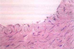
Fig. 3-7. LM of a muscular artery. These arteries have an internal elastic lamina made up of elastic fibers produced by fibroblasts and smooth muscle cells. The lamina is located near the lumen where, in fixed tissue, it has a folded, ribbon-like configuration and is glassy or translucent (hyaline) in appearance. (H&E; X250.).
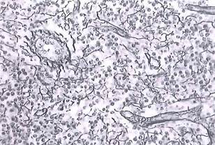
Fig. 3-8. LM of a lymph node permitting visualization of the reticular fibers produced by reticular cells (compare to Fig. 3-6). Note the arrangement of reticular fibers around smooth muscle cells in small blood vessels. (Silver stain; X 250.).
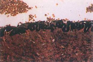
Fig. 3-9. LM of a muscular artery. The internal elastic lamina is selectively stained black. Compare it with the image directly above. (Verhoeff stain) (X 250.).
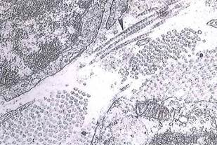
Fig. 3-10. ТЕМ of the extracellular matrix subtending epithelial cells and basal lamina. Reticular fibers are generally smaller in diameter than collagen fibers but also possess 64 to 67 nm periodicity. Observe reticular fibers in longitudinal section adjacent Ъ to the basal lamina (arrowhead), and compare them with the cross sections of larger collagen fibers seen in the bundles below. The basal lamina of an epithelial cell (upper left) is composed of collagen type IV. (x 11,000.).
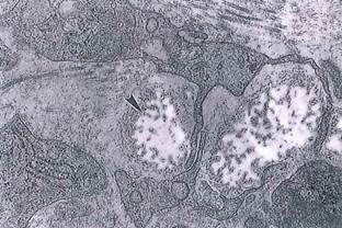
Fig. 3-11. Higher-magnification ТЕМ showing the microfibrillar component (arrowhead) of elastic fibers and their arrangement around amorphous elastin (nonstaining in this fixation). Collagen fibers are also seen, (x 9,500.).
Date added: 2022-12-11; views: 1030;