Erythrocytes and platelet seen by ТЕМ. ТЕМ of lymphocyte
Fig. 6-1. LM of red blood cells, platelets (arrowheads), and a lymphocyte in peripheral blood. Note thin, lightly basophilic rim of cytoplasm surrounding nucleus of lymphocyte. (Wright's stain; x 1,100.).
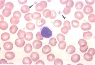
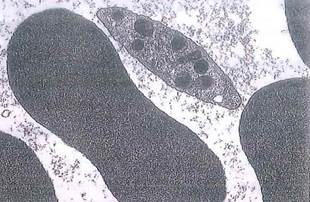
Fig. 6-2. Erythrocytes and platelet seen by ТЕМ. Erythrocyte has dense, homogeneous appearance because of presence of hemoglobin and absence of cell organelles. Locate cisternae of platelet's dense tubular system, dense granules, and microtubules. Compare platelet to those seen in Fig. 6-1 by LM. (X 9,000.).
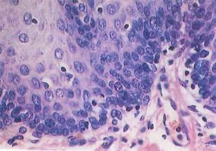
Fig. 6-3. LM of esophageal mucosa. Lymphocytes are identified by small, dense nuclei lying between epithelial cells and within underlying connective tissue. (H&E; X250.).
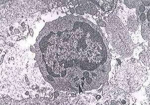
Fig. 6-4. ТЕМ of lymphocyte. Membranous organelles are few, but there are abundant free ribosomes in surrounding cytoplasm. A pair of centrioles (arrowhead) and several mitochondria are seen, (x 5,100.).
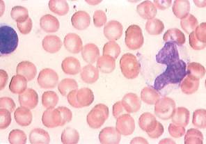
Fig. 6-5. LM of peripheral blood. Compare monocyte (right) with lymphocyte (left) and red blood cells, which are scattered throughout field. Monocyte is a large cell with gray, vacuolated cytoplasm, a few azure granules, and indented nucleus in which chromatin has a "raked" appearance. In contrast, the smaller lymphocyte possesses condensed chromatin and is surrounded by thin rim of basophilic cytoplasm. (Wright’s stain; x 1,100.).
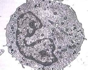
Fig. 6-6. Monocyte (ТЕМ) exhibiting small lysosomal granules (azure granules) and indented nucleus. Monocytes are precursors of tissue macrophages, g, Golgi; c, centriole. (x 6,500.).
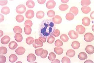
Fig. 6-7. LM of polymorphonuclear leukocyte (also known as PMN or neutrophil) in peripheral blood. Notice multilobed nucleus and azure and specific granules. Compare in size with red blood cells. (Wright’s stain; X 1,100.).
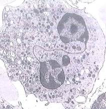
Fig. 6-8. ТЕМ of polymorphonuclear leukocyte. Observe multilobed nucleus and specific granulation. (X 5,200.).
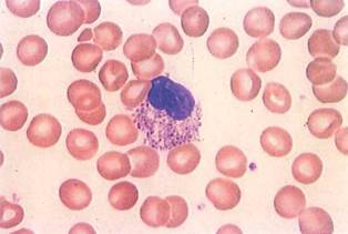
Fig. 6-9. LM of eosinophil with a bilobed nucleus in peripheral blood. Specific granules are large and acidophilic. (Wright’s stain; X 1,100.).
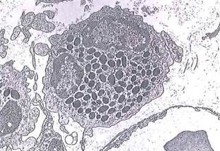
Fig. 6-10. ТЕМ of eosinophil adjacent to capillary in connective tissue. Specific granules are large and electron-dense and usually contain angular crystalloid. Nucleus is bilobed. (Х5,100.).
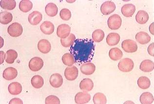
Fig. 6-11. LM of basophil in peripheral blood. Cytoplasm of mature basophil is clear and contains numerous large, basophilic specific granules, which usually overlie and obscure bilobed nucleus. (Wright’s stain; X 1,100.).
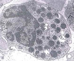
Fig. 6-12. ТЕМ of human basophil. Specific granules are large and regular in size and shape and contain histamine, heparin, SRS (slow reacting substance), and serotonin, which are powerful vasoactive mediators. Granules are often disrupted owing to difficulty in fixing tissue, (x 10,500.).
Date added: 2022-12-11; views: 917;