High-resolution SEM of the cytosolic surface of the RER. Diagram of the pathways of the lysosomal system
Fig. 1-21. ТЕМ of liver cell cytoplasm showing an array of parallel cisternae of the RER and several adjacent lysosomes containing dense whorls of membranous elements called myelin figures, (x 83,300.).
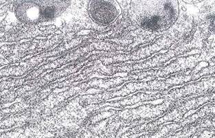
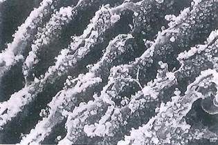
Fig. 1-22. SEM of cisternae of the RER. Ribosomes, seen attached to the cytosolic surface of the endoplasmic reticulum, transcribe mRNA as they synthesize proteins which are then sequestered within the cisternae of the RER before transfer to the Golgi apparatus for packaging. (X 20,500.).

Fig. 1-23. High-resolution SEM of the cytosolic surface of the RER. Ribosomes and polyribosomes (linked by messenger RNA, arrowheads) can be seen on the surface of the cisternae. (x 176,000.).

Fig. 1-24. ТЕМ of a centriole showing the triplet arrangement of microtubules. Electron-dense pericentriolar satellites (pc) serve as nucleation sites for cytoplasmic microtubules (mt). Cytoplasmic filaments and polyribosomes are also visible. (X 87,000.).
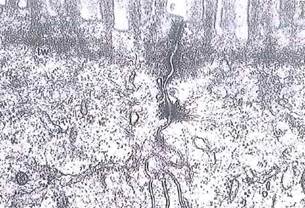
Fig. 1-25. ТЕМ of the adluminal portions of two polarized intestinal epithelial cells showing the junctional complex (called terminal bar by LM) and the microfilaments of the terminal web (tw) just deep to the microvillous border. Note the desmosome with tonofilaments (keratin type of intermediate filaments) extending into the cytoplasm of each cell. Lateral borders of the cells interdigitate. (X 36,000.).
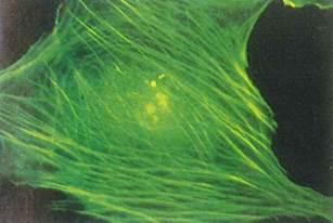
Fig. 1-26. An immunofluorescent micrograph demonstrating the localization of actin within NRK tissue culture cells. Bundles of actin filaments, sometimes called stress fibers, can be seen coursing in a diagonal pattern through the cytoplasm and around the periphery of the cell, (x 500.).
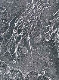
Fig. 1-27. ТЕМ of a deep etch preparation of a fibroblast cytoskeleton showing several actin cables (stress fibers; see Fig. 126) attached to the cytoplasmic surface of the plasma membrane. The actin filaments have been decorated with the SI fragment of myosin, resulting in the filaments having a barbed appearance. Coated vesicles, involved in receptor-mediated endocytosis, can be seen as honeycomb-shaped structures associated with the cytoplasmic surface of the membrane, (x 52,500.).
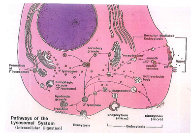
Fig. 1-28. Diagram of the pathways of the lysosomal system involved with endocytic uptake and intracellular digestion. Endocytic uptake or heterophagy includes phagocytosis and intracellular degradation of exogenous particles such as bacteria (see Figs. 1-29 and 1-30) as well as pinocytosis of fluid. Autophagy involves the degradation of endogenous intracellular organelles within lysosomal vacuoles (see Figs. 1-31 and 1-32). Receptor-mediated endocytosis involves the selective rapid uptake of molecules (hormones, growth factors, cholesterol, iron, etc.) from the extracellular fluid by specific protein receptors on the extracellular surface of the cell membrane (see Figs. 1-33 and 1-34). Following ingestion via clathrin-coated vesicles, recepto- somes (endosomes) carry the ligand-receptor complexes into the cell where they may be delivered to the lysosomal system. In some instances, the receptors may be dissociated from the ingested ligand and recycled to the cell surface.

Fig. 1-29. Acridine orange stain of peritoneal exudate cells showing the phagocytosis of rod-shaped bacteria by a macrophage. Endocytosed bacteria in the phagolysosomal system that are dead stain orange, while viable bacteria are green. (X700.).

Fig. 1-30. SEM of peritoneal macrophage showing both membrane-adherent and partially ingested bacteria. Note the close adhesion of the macrophage membrane to the bacterium being endocytosed. (Х 1000.).
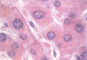
Fig. 1-31. LM of liver cell. Observe the golden brown lipofuscin pigment, which represents residual bodies (lysosomes) in the liver cell cytoplasm. (H&E; X 850.).
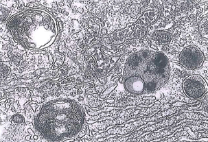
Fig. 1-32. ТЕМ of liver cell cytoplasm containing several residual bodies (lysosomes) with undigested cytoplasmic residues scattered between elements of the RER and Golgi apparatus. The latter can be identified by the presence of lipoprotein particles within the lumen of the cisternae. (X 32,200.).

Fig. 1-33. ТЕМ of receptor-mediated endocytosis in cultured human KB cells. A, A colloidal gold-labeled antitransferrin receptor antibody was used to localize the transferrin receptor within a clathrin-coated pit. B, After clustering of receptors, vesicles bud off from the surface and form receptosomes or endosomes, a process requiring about 20 seconds. The pH of the receptosome decreases, dissociating the ligand from the receptor and allowing the receptor to recycle to the cell surface, while the ligand eventually becomes degraded in the lysosomal apparatus. (x 81,500.).
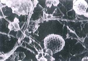
Fig. 1-34. SEM of the interior of a cell showing several different sizes of filaments of the cytoskeleton and clathrin-coated pit and vesicles. The coated pit (center right) has not yet detached from the plasmalemma, (x 82,000.).
Date added: 2022-12-11; views: 1173;