Acquisition of Medical Images
A biomedical image analysis system comprises three major elements: an image acquisition system, a computer for processing the acquired information, and a display system for visualizing the processed images. In medical image acquisition, the primary objective is to capture and record information about the physical and possibly functional properties of organs or tissues by using either external or internal energy sources or a combination of these energy sources.
Conventional Radiography. In conventional radiography, a beam of x rays from an external source passing through the body is differentially absorbed and scattered by structures. The amount of absorption depends on the composition of these structures and on the energy of the x ray beam.
Conventional imaging methods, which are still the most commonly used diagnostic imaging procedure, form a projection image on standard radiographic film. With the advent of digital imaging technology, radiographs, which are x-ray projections, are increasingly being viewed, stored, transported, and manipulated digitally. Figure 1 shows a normal chest x-ray image.
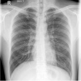
Figure 1. X-ray image of a chest
Computed Tomography. The realization that x-ray images taken at different angles contain sufficient information for uniquely determining the internal structures, led to the development of x-ray CT scanners in the 1970s that essentially reconstruct accurate cross-sectional images from x-ray radiographs.
The conventional x-ray CT consists of a rotating frame that has an x-ray source at one end and an array of detectors that accurately measure the total attenuation along the path of the x ray at the other end. A fan beam of x rays is created as the rotating frame spins the x-ray tube and detectors around the patient. During the 360° rotation, the detector captures numerous snapshots of the attenuated x-ray beam corresponding to a single slice of tissue whose thickness is determined by the collimation of the x-ray beam.
This information is then processed by a computer to generate a 2-D image of the slice. Multiple slices are obtained by moving the patient in incremental steps. In the more recent spiral CT (also known as the helical CT), projection acquisition is carried out in a spiral trajectory as the patient continuously moves through the scanner. This process results in faster scans and higher definition of internal structures, which enables greater visualization of blood vessels and internal tissues.
CT images of a normal liver and brain are shown in Fig. 2. In comparison with conventional x-ray imaging, CT imaging is a major breakthrough. It can image the structures with subtle differences in x-ray absorption capacity even when almost obscured by a structure with a strong ability on x-radiation absorption. For example, the CT can image the internal structures of the brain, which is enclosed by the skull [as shown in Fig. 2(b)], whereas the x ray fails to do so.
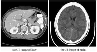
Figure 2. CT images of normal liver and brain
Magnetic Resonance Imaging. Medical imaging methods using magnetic resonance include MRI, PET, and SPECT. MRI is based on the principles of nuclear magnetic resonance (NMR), which is a spectroscopic technique used to obtain microscopic chemical and physical information about molecules. An MRI can produce high-quality multidimensional images of the inside of the human body, as shown in Fig. 3, providing both structural and physiologic information of internal organs and tissues.
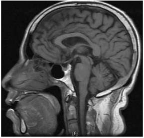
Figure 3. MRI of the brain
Unlike the CT, which depicts the x- ray opacity of the structure being imaged, MRIs depict the density as well as biochemical properties based on physiologic function, including blood flow and oxygenation. A major advantage of the MRI is the fast signal acquisition with a very high spatial resolution.
Radiographic imaging modalities such as those based on x rays provide anatomical information about the body but not the functional or metabolic information about an organ or tissue. In addition to anatomical information, MRI methods are capable of providing some functional and metabolic information.
Nuclear medicine-based imaging systems image the distribution of radioisotopes distributed within specific organs of interest by injection or inhalation of radio-pharmaceuticals that metabolize the tissue, which makes them a source of radiation. The images acquired by these systems provide a direct representation of the metabolism or function of the tissue or organ being imaged as it becomes a source of radiation that is used in the imaging process. SPECT and PET are nuclear medicine-based imaging systems.
SPECT systems use gamma cameras to image photons that are emitted during decay. Like the x-ray CT, many SPECT systems rotate a gamma camera around the object being imaged and process the acquired projections using a tomographic reconstruction algorithm to yield a 3-D reconstruction. SPECT systems do not provide good resolution images of anatomical structures like CT or MR images, but they show distribution of radioactivity in the tissue, which represents a specific metabolism or blood flow as shown in Fig. 4(a).
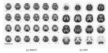
Figure 4. SPECT and PET sequences of a normal brain
PET systems, like SPECT, also produce images of the body by detecting the emitted radiation, but the radioactive substances used in PET scans are increasingly used with CT or MRI scans so as to provide both anatomical and metabolic information. Some slices of a PET brain image are shown in Fig. 4(b).
Ultrasound Imaging. Ultrasound or acoustic imaging is an external source-based imaging method. Ultrasound imaging produces images of organs and tissues by using the absorption and reflection of ultrasound waves traveling through the body (Fig. 5).
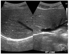
Figure 5. Ultrasound image of a normal liver
It has been successfully used for imaging of anatomical structures, blood flow measurements, and tissue characterization. A major advantage of this method, which does not involve electromagnetic radiation, is that it is almost non nonintrusive, and hence, the examined structures can be subjected to uninterrupted and long-term observations while the subject does not suffer any ill effects.
Date added: 2024-03-07; views: 686;