Medical Image Registration
Multiple images of the same subject, acquired possibly using different medical imaging modalities, contain useful information usually of complementary nature. Proper integration of the data and tools for visualizing the combined information offers potential benefits to physicians. For this integration to be achieved, there is a need to spatially align the separately acquired images, and this process is called image registration.
Registration involves determining a transformation that can relate the position of features in one image with the position of the corresponding features in another image. To determine the transformation, which is also known as spatial mapping, registration algorithms use geometrical features such as points, lines, and surfaces that correspond to the same physical entity visible in both images.
After accurate registration, different images will have the same coordinate system so that each set of points in one image will occupy the same volume as the corresponding set of points in another image. In addition to combining images of the same subject from different modalities, the other applications of image registration include aligning temporal image sequences to compensate for motion of the subject between scans and image guidance during medical procedures.
The evaluation of the transformation parameters can be computationally intensive but can be simplified by assuming that the structures of interest do not deform or distort between image acquisitions. However, many organs do deform during image acquisition, for example, during the cardiac and respiratory cycles. Many medical image registration algorithms calculate rigid body or affine transformations, and thus, their applicability is restricted to parts of the body where deformation is small.
As bones are rigid, rigid body registration is widely used where the structures of interest are either bone or are enclosed in bone. The brain, which is enclosed by the skull, is reasonably nondeformable, and several registration approaches have been applied to the rigid body registration of brain images. Figure 11 shows an example of rigid registration of brain MR images. Image registration based on rigid body transformations is widely used for aligning multiple 3-D tomographic images of the same subject acquired using different modalities (intermodality registration) or using the same modality (intramodality registration).
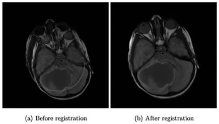
Figure 11. (a) Result of direct overlapping of brain MR images without registration. (b) Result of overlapping of images after rigid registration
The problem of aligning images of structures with deformed shapes is an active area of research. Such deformable (nonaffine) registration algorithms usually involve the use of an initial rigid body or affine transformation to provide a starting estimate.
After registration, fusion is required for the integrated display. Some fusion methods involve direct combination of multiple registered images. One example is the superimposition of PET data on MRI data to provide a single image containing both structure and function. Another example involves the use of MR and CT combined to delineate tumor tissues.
Atlas-Based Approaches. In atlas-based registration and segmentation of medical images, prior anatomical and/or functional knowledge is exploited. The atlas is actually a reference image in which objects of interest have been carefully segmented. In this method , the objective is essentially to carry out a nonrigid registration between the image of the patient and the atlas. The first step, known as atlas warping, involves finding a transformation that maps the atlas image to the target image to be segmented.
The warping usually consists of a combination of linear and nonlinear transformations to ensure good registration despite anatomical variability. Even with nonlinear registration methods, accurate atlas-based segmentation of complex structures is difficult because of anatomical variability, but these approaches are generally suited for segmentation of structures that are stable in large numbers of people. Probabilistic atlases help to overcome anatomical variability.
Date added: 2024-03-07; views: 634;