History of Mineralogy. The Second Part
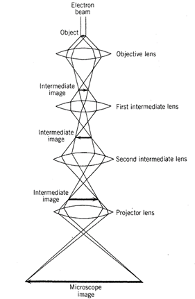
Since early 1970 another electron beam instrument, which can magnify the internal architecture of minerals many millions of times, has produced elegant and powerful visual images of atomic structures. This instrument, the transmission electron microscope, is illustrated in Fig. 1.12, and a schematic representation of the magnification process of an object into a final and highly enlarged image is shown in Fig. 1.13.

FIG. 1.12. Transmission electron microscope, Philips Tecnai ТЕМ manufactured by the FEI Company. The tall vertical feature is the electron column with control mechanisms on either side and a viewing screen at table-top level; the viewing screen is used for transmission electron microscope (ТЕМ) imaging and for display of electron diffraction patterns. In the middle of the electron column and slightly to the right is the sample assembly holder.
An energy dispersive X-ray detector (EDX) with liquid nitrogen dewar for cooling is affixed to the right of the column. Computer control of the whole system and a monitor for data display are to the far right. (Courtesy FEI Company, Hillsboro, Or.)

FIG. 1.13. Schematic cross section through the column of a transmission electron microscope, showing the electron beam path for structural imaging. The four lenses are electromagnetic lenses
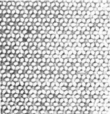
The most visually instructive application of this technique is known as high-resolution transmission electron microscopy (HRTEM), which allows the study of crystalline materials at resolutions approaching the scale of atomic distances (see P. R. Buseck 1983). The technique can produce projected two-dimensional images of three-dimensional crystalline structures. These images show that many minerals have infinitely extending, periodic (meaning: perfectly repeating) internal structural arrangements; an example of such a "perfect" structure is illustrated by the HRTEM image in Fig. 1.14 for the chemically complex mineral tourmaline. The HRTEM images have also shown that minerals may contain defects that are deviations from idealized ("perfect") structures (see Figs. 4.53 and 4.54).

FIG. 1.14. High-resolution transmission electron microscope (HRTEM) image of the structure of tourmaline. (From lijima, S., Cowley, J. M., and Donnay, G., 1973, High-resolution electron microscopy of tourmaline crystals. Tschermaks Mineralogische Petrographische Mitteilungen, v. 20, pp. 216-224.) The white areas in the photograph correspond to regions of low electron density in the structure of tourmaline. The six-fold pattern is the image of the six-fold Si6018 ring in tourmaline (compare with Fig. 11.14). The bar scale represents 15 angstroms
The field of mineral science now encompasses a very broad area of study that includes X-ray, electron, and neutron diffraction by minerals, mineral synthesis, crystal physics, the evaluation of the thermodynamic stability of minerals, petrography (the study of rocks and minerals in thin section), petrology (the study of rocks), experimental petrology, and aspects of metallurgy and ceramics.
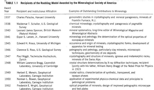
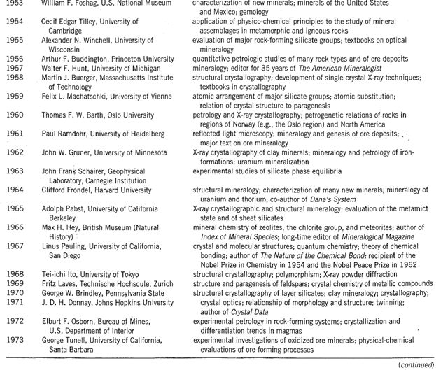
Because it is difficult to predict which contributions (made in relatively recent times) to the science of mineralogy will prove to be most enduring and important, Table 1.1 provides a tabulation of the Roebling medalists and some of their major research contributions. This list illustrates the diversity of internationally recognized professional contributions, and it is a reasonable assumption that it includes those mineralogists whom future historians will regard as the mineralogical giants of our time.


The Roebling medal was established in 1937 by the Mineralogical Society of America in memory of Colonel Washington A. Roebling (1837-1926), who had made a generous financial gift to the society in 1926. Colonel. Roebling, the designer of such well- known suspension bridges as those over the Niagara River at Niagara Falls, over the Allegheny River at Pittsburgh, over the Ohio River in Cincinnati, and the Brooklyn Bridge over the East River in New York City, had a deep, life-long interest in the study of minerals. The Roebling medal signifies the highest recognition of achievement American mineralogy can bestow on outstanding investigators in the United States or abroad. The presentation and acceptance speeches for each Roebling award can be found in American Mineralogist.
In 1927, Roebling's son John donated his father's mineral collection of about 16,000 specimens to the National Museum of Natural History (Smithsonian Institution) in Washington, D.C. This collection, known as the Washington A. Roebling mineral collection, was undoubtedly one of the largest and finest private collections of its time. This acquisition, together with another collection of about 9100 specimens (the Canfield collection), made the Smithsonian mineral collection one of the best in the world (A. Roe 1990, Washington A. Roebling, his life and his mineral collection, Mineralogical Record, v. 21, pp. 13-30).
Date added: 2022-12-31; views: 630;
