The plasma membrane
The existence of the plasma membrane (or cell membrane or plasmalemma) was indirectly demonstrated in the middle of the 19th century by Karl Wilhelm von Nägeli (Kilchberg 1817 - Munich 1891), who added dye to a container containing cells in a solution of water and salts.
He observed that the cells did not or only faintly took up the color, while the whole solution was uniformly colored (Figure 2.7). He also observed that a cell placed in a diluted solution swelled due to water ingress, while when placed in a concentrated solution, it wrinkled due to water loss.
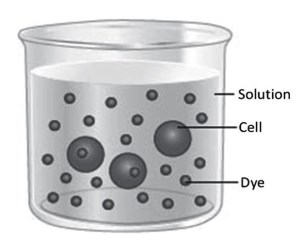
Figure 2.7. The dye is spread evenly in the solution but not in the cells
Von Nägeli, therefore, had observed a series of phenomena, the coloring of the cytoplasm, the swelling and the wrinkling, that led him to suppose the existence of a barrier between the inside and the outside of the cell, but he was unable to see it. Only with the use of the electron microscope did J. David Robertson (1955) discover that the cells were surrounded by a three-layer membrane about 10 nm thick.
von Nägeli's experiments indirectly demonstrated the existence of the plasma membrane, but also two of its fundamental functions. The first is the barrier function, which keeps the cell's internal environment separate from the outside and prevents the entry of substances unsuitable for its operation, such as dye, and the uncontrolled escape of useful substances. This property is due to the composition of the plasma membrane which makes it impermeable to water-soluble molecules.
The experiment with concentrated or diluted solutions, on the other hand, demonstrates the selective function of the membrane. In fact, with both diluted and concentrated solutions, only water moves through the plasma membrane and not the solutes present inside and outside the cell. This means that the plasma membrane is able to select which molecules can pass through it, generally thanks to specific proteins present on the membrane itself.
The proteins present on the external face of the plasma membrane can also be receptors for hormones and neurotransmitters, making the membrane itself perform the function of communication between the outside and inside of the cell. Numerous protein molecules with enzymatic functions can function correctly because they are in an orderly sequence on the internal face of the plasma membrane, such as the protein sequence

thus performing an important biochemical function.
The widely-accepted plasma membrane model is the double phospholipid fluid mosaic layer (Singer and Nicolson, 1972). If we imagine looking at a cell from the outside, we observe a heterogeneous mosaic of single molecules, or small groups of molecules, of a protein nature, distributed more or less uniformly within the phospholipid double layer. It has been calculated that an average of 106 phospholipid molecules per μm2 of membrane and as many protein molecules are present.
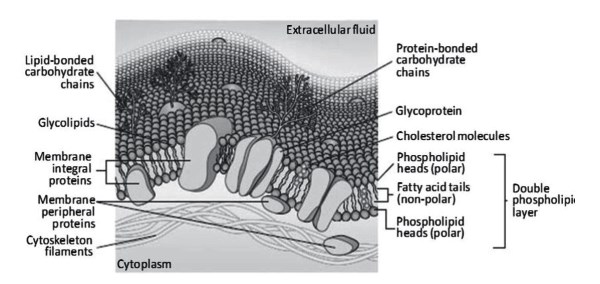
Figure 2.8. The plasma membrane consists mainly of phospholipids and proteins
The fluidity of the membrane is due to the fact that the forces holding the tails of the molecules in the two layers and those of each layer together are the relatively weak van der Waals forces. These forces allow phospholipid molecules to move rarely, on average once every thirty days, from one layer to the other and more frequently, it is believed, about 107 times per second, within a layer. It is because of this fluidity that the membrane can repair small tears that may form.
The plasma membrane consists almost entirely of two types of molecules: phospholipids and proteins (Figure 2.8).
Date added: 2024-07-10; views: 622;