
CNS Protection: Meninges, CSF, and Blood-Brain Barrier
The central nervous system (CNS)—comprising the brain within the skull and the spinal cord within the vertebral column—requires specialized protection from bony surfaces and potentially harmful bloodborne substances. This protection is provided by three membranous layers called the meninges: the thick dura mater adjacent to the bone, the middle arachnoid mater, and the thin pia mater directly covering the neural tissue (Figure 6.47). The subarachnoid space, located between the arachnoid mater and pia mater, is filled with cerebrospinal fluid (CSF). The meninges and their associated structures not only support and protect the CNS but also facilitate CSF circulation and absorption. Meningitis, an infection of the meninges within the subarachnoid space, can lead to elevated intracranial pressure, seizures, and loss of consciousness.
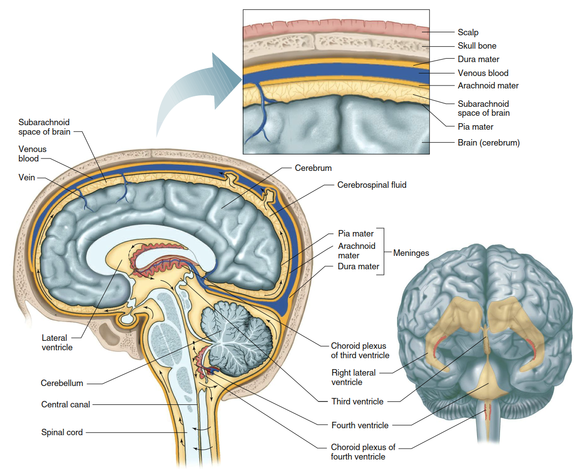
Figure 6.47. The four interconnected ventricles of the brain. The lateral ventricles form the first two. The choroid plexus forms the cerebrospinal fluid (CSF), which flows out of the ventricular system at the brainstem (arrows)
CSF production occurs via a specialized epithelial structure known as the choroid plexus, which is composed of ependymal cells. This plexus produces CSF at a rate that completely replenishes the entire fluid volume approximately three times per day. The black arrows in Figure 6.47 illustrate the directional flow of CSF, which circulates through the interconnected ventricular system toward the brainstem. From there, CSF passes through small openings into the subarachnoid space surrounding both the brain and spinal cord. Assisted by pressure changes from circulation, respiration, and body posture, CSF ultimately flows to the superior aspect of the brain’s outer surface, where most of it reenters the bloodstream via one‑way valves within large veins.
CSF analysis provides critical diagnostic information for neurological diseases, including meningitis. A CSF sample is typically obtained by inserting a large needle into the spinal canal below the second lumbar vertebra, the level at which the spinal cord ends (see Figure 6.42). Consequently, the CNS effectively “floats” within a cushion of CSF, which absorbs sudden mechanical shocks and protects the soft neural tissues from jarring movements. If CSF outflow becomes obstructed, fluid accumulates, resulting in hydrocephalus (“water on the brain”). In severe, untreated hydrocephalus, the resulting intraventricular pressure compresses cerebral blood vessels, potentially leading to inadequate neuronal blood flow, neuronal damage, and cognitive dysfunction.
Although some evidence suggests that CSF may serve limited nutritive functions, the brain—like all tissues—derives its nutrients from the blood. Under normal conditions, glucose is the brain’s sole metabolic substrate for energy production, with most energy from oxidative glucose metabolism transferred to ATP. The brain possesses negligible glycogen stores, making it entirely dependent on a continuous blood supply of glucose and oxygen. The most common form of brain damage results from reduced regional blood flow: when neurons are deprived of blood supply for even a few minutes, they cease functioning and undergo cell death. Such neuronal death caused by vascular disease is termed a stroke. Despite constituting only 2% of total body weight, the adult brain receives 12–15% of the cardiac output, supporting its exceptionally high oxygen utilization. When regional cerebral blood flow falls to 10–25% of normal levels, energy‑dependent membrane ion pumps fail, ion gradients diminish, extracellular potassium concentration rises, and membranes depolarize.
The blood‑brain barrier (BBB) fundamentally distinguishes CNS exchange from the relatively unrestricted diffusion of non‑protein substances seen in other organs. A complex ensemble of BBB mechanisms tightly regulates which substances enter the brain’s extracellular fluid and the rates at which they do so. These mechanisms minimize the access of many harmful agents to neurons, but they also limit the entry of potentially beneficial therapeutic drugs. Anatomically, the BBB is formed by cells lining the smallest cerebral blood vessels, featuring tight junctions and specialized physiological transport systems that handle different classes of molecules distinctly. Substances that readily dissolve in the lipid components of plasma membranes enter the brain rapidly; therefore, the extracellular fluid of the brain and spinal cord is a product of—yet chemically distinct from—blood.
The BBB also explains certain drug actions, as illustrated by the morphine‑heroin comparison. Morphine and heroin differ chemically only slightly: morphine possesses two hydroxyl groups, whereas heroin has two acetyl groups (—COCH₃). This difference makes heroin more lipid‑soluble, allowing it to cross the BBB more readily than morphine. However, once heroin enters the brain, enzymes remove its acetyl groups, converting it into morphine. The resulting morphine, being less lipid‑soluble, becomes effectively trapped within the brain, where it can exert prolonged effects. Other drugs with rapid CNS actions due to high lipid solubility include barbiturates, nicotine, caffeine, and alcohol.
Many substances that do not dissolve readily in lipids—such as glucose and other essential brain metabolites—nevertheless enter the brain quickly via membrane transport proteins located on cells lining the smallest cerebral vessels. Similar transport systems actively move substances out of the brain into the blood, preventing the accumulation of molecules that could interfere with neuronal function. A protective barrier also exists between blood in the choroid plexus capillaries and the CSF, making CSF a selective secretion. For example, potassium (K⁺) and calcium (Ca²⁺) concentrations are slightly lower in CSF than in plasma, whereas sodium (Na⁺) and chloride (Cl⁻) concentrations are slightly higher. The choroid plexus vessel walls exhibit limited permeability to toxic heavy metals such as lead, thereby affording additional protection to the brain.
Over time, the CSF and the extracellular fluid of the CNS reach diffusion equilibrium. Thus, the restrictive, selective barrier mechanisms in the cerebral capillaries and choroid plexuses collectively regulate the extracellular environment surrounding the neurons of the brain and spinal cord. This integrated system of meninges, CSF dynamics, and blood‑brain barrier ensures both mechanical cushioning and biochemical stability, which are essential for normal CNS function.
Date added: 2026-07-14; views: 141;