
Synaptic Integration in Neurons: EPSP, IPSP, Summation, and Threshold
In most neurons, a single excitatory synaptic event is insufficient to reach the threshold of the postsynaptic neuron. For example, one excitatory postsynaptic potential (EPSP) may be only 0.5 mV, whereas a depolarization of approximately 15 mV is required to bring the membrane to threshold. Therefore, an action potential can only be initiated through the combined effects of many simultaneously active excitatory synapses.
Out of the thousands of synapses on a typical neuron, hundreds are likely active at the same time or within a sufficiently short interval for their effects to add together. The membrane potential of the postsynaptic neuron at any moment is thus the result of all synaptic activity affecting it at that instant. When excitatory input predominates, the membrane depolarizes toward threshold; when inhibitory input predominates, either a hyperpolarization or a stabilization of the membrane potential occurs.
A simple experiment (Figure 6.31) demonstrates how EPSPs and inhibitory postsynaptic potentials (IPSPs) interact. Assume three synaptic inputs to the postsynaptic cell: axons A and B provide excitatory synapses, while axon C provides an inhibitory synapse. Stimulators on axons A, B, and C allow individual activation, and an electrode in the cell body records the membrane potential. In part 1 of the experiment, stimulating axon A twice in succession shows no interaction between the two EPSPs because the membrane potential change from an EPSP is short‑lived, like all graded potentials; within a few milliseconds, the cell returns to rest.
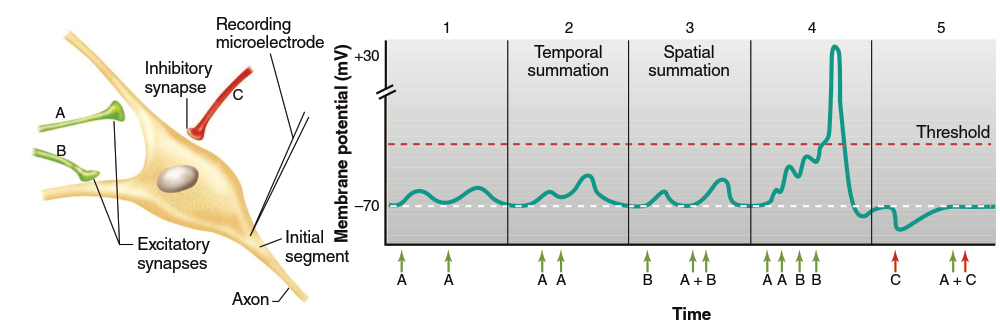
Figure 6.31. Interaction of EPSPs and IPSPs at the postsynaptic neuron. Presynaptic neurons (A–C) were stimulated at times indicated by the arrows, and the resulting membrane potential was recorded in the postsynaptic cell by a recording microelectrode
In part 2, the second stimulation of axon A occurs before the first EPSP has decayed, causing the second synaptic potential to add to the previous one and produce a greater depolarization. This phenomenon is called temporal summation, as the input signals arrive from the same presynaptic cell at different times. Summation happens because an additional influx of positive ions enters before ions leaking out through the membrane have restored the resting potential.
Part 3 of Figure 6.31 first stimulates axon B alone to determine its response, then simultaneously stimulates axons A and B. The EPSPs from two separate neurons also add together in the postsynaptic neuron, resulting in a larger depolarization. Although the two stimulations must occur close in time for summation to occur, this is termed spatial summation because the inputs originate at different locations on the cell. Through spatial and temporal summation, multiple EPSPs can increase the inward flow of positive ions and bring the postsynaptic membrane to threshold, thereby initiating action potentials (see part 4 of Figure 6.31).
So far, only patterns of excitatory synapse interaction have been tested. Because EPSPs and IPSPs arise from oppositely directed local currents, they tend to cancel each other, producing little or no net change in membrane potential when both A and C are stimulated (Figure 6.31, part 5). Inhibitory potentials can also exhibit spatial and temporal summation.
Depending on the postsynaptic membrane resistance and the amount of charge moving through ligand‑gated ion channels, the synaptic potential spreads to a greater or lesser degree across the plasma membrane. During activation of an excitatory synapse, a large membrane area becomes slightly depolarized; during activation of an inhibitory synapse, it becomes slightly hyperpolarized or stabilized, although these graded potentials decrease with distance from the synaptic junction (Figure 6.32). Inputs from multiple synapses can summate, potentially triggering an action potential.
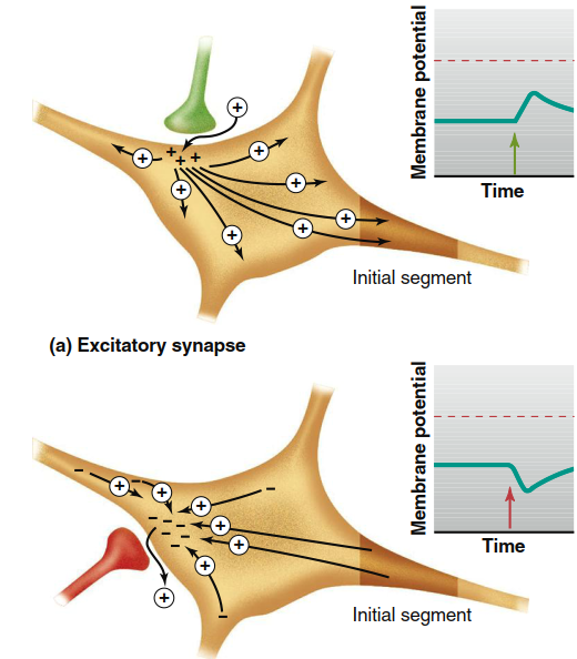
Figure 6.32. Comparison of excitatory and inhibitory synapses, showing current direction through the postsynaptic cell following synaptic activation. (a) Current through the postsynaptic cell is away from the excitatory synapse and may depolarize the initial segment. (b) Current through the postsynaptic cell is toward the inhibitory synapse and may hyperpolarize the initial segment. The arrow on the graph indicates moment of stimulus
The previous examples referred to the threshold of the postsynaptic neuron as if it were uniform across the cell, but different parts have different thresholds. In general, the initial segment has a more negative threshold (i.e., much closer to the resting potential) than the membrane of the cell body and dendrites. This lower threshold is due to a higher density of voltage‑gated Na⁺ channels in that area. Consequently, the initial segment is most responsive to small changes in membrane potential arising from synaptic inputs on the cell body and dendrites. When enough EPSPs summate, the initial segment reaches threshold, and the resulting action potential propagates from this point down the axon.
The fact that the initial segment usually has the lowest threshold explains why the location of individual synapses on the postsynaptic cell matters. A synapse located near the initial segment produces a greater voltage change there than a synapse on the outermost branch of a dendrite, because it exposes the initial segment to a larger local current. However, in some neurons, signals from dendrites far from the initial segment may be boosted by the presence of voltage‑gated Na⁺ channels in those dendritic regions.
Postsynaptic potentials last much longer than action potentials. If cumulative EPSPs keep the initial segment depolarized to threshold after an action potential has fired and the refractory period ends, a second action potential will occur. In fact, as long as the membrane remains depolarized to threshold, action potentials continue to arise. Therefore, neuronal responses almost always occur in bursts of action potentials rather than as single, isolated events.
Date added: 2026-07-14; views: 147;