
Synaptic Transmission: Electrical and Chemical Synapses in the Nervous System
A synapse is an anatomically specialized junction between two neurons, where electrical activity in a presynaptic neuron influences the electrical activity of a postsynaptic neuron. Anatomically, synapses consist of parts of the presynaptic and postsynaptic neurons along with the extracellular space between these two cells. Recent estimates indicate that the human central nervous system (CNS) contains over 10¹⁴ (100 trillion) synapses.
Activity at synapses can either increase or decrease the likelihood that the postsynaptic neuron will fire action potentials by producing brief, graded potentials in the postsynaptic membrane. At an excitatory synapse, the membrane potential of a postsynaptic neuron is depolarized, bringing it closer to the threshold for action potential generation. Conversely, at an inhibitory synapse, the membrane is hyperpolarized (driven farther from threshold) or stabilized at its resting potential, reducing neuronal excitability.
Hundreds or thousands of synapses from different presynaptic cells can converge onto a single postsynaptic cell, a phenomenon known as convergence (Figure 6.25). Conversely, a single presynaptic cell can send branched axons to affect many postsynaptic cells, termed divergence (Figure 6.25). Convergence allows information from multiple sources to influence a cell’s activity, while divergence enables one cell to modulate multiple neural pathways.
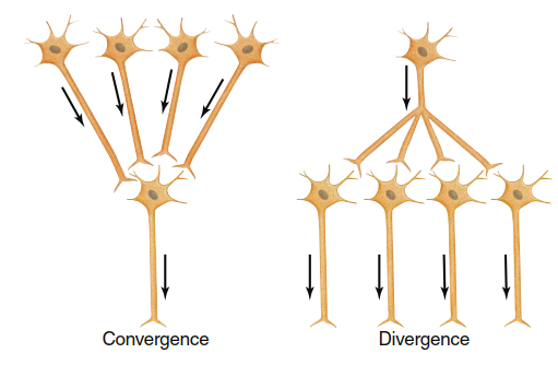
Figure 6.25. Convergence of neural input from many neurons onto a single neuron, and divergence of output from a single neuron onto many others. Arrows indicate the direction of transmission of neural activity
The excitability level of a postsynaptic cell at any given moment—how close its membrane potential is to threshold—depends on the number of active synapses and the balance between excitatory and inhibitory inputs. If the postsynaptic membrane reaches threshold, it generates action potentials that propagate along its axon to the axon terminals. These terminals subsequently influence the excitability of other cells, continuing the signal transmission cascade.
Functional Anatomy of Synapses. There are two primary types of synapses: electrical synapses and chemical synapses.
Electrical Synapses. At electrical synapses, the plasma membranes of the presynaptic and postsynaptic cells are connected by gap junctions (Figure 6.26a; see also Figure 3.9). These gap junctions allow local currents generated by arriving action potentials to flow directly through connecting channels from one neuron to the next. This direct current flow depolarizes the membrane of the postsynaptic neuron to threshold, continuing action potential propagation. A key advantage of electrical synapses is extremely rapid cell-to-cell communication, with no delay from chemical messenger release.
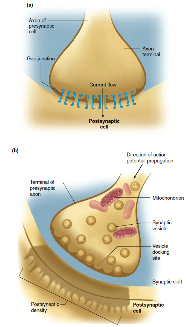
Figure 6.26. (a) An electrical synapse. Note that there is very little space between the two cells, which are connected by gap junctions through which ions diffuse. (b) Diagram of a chemical synapse. Some vesicles are docked at the presynaptic membrane, ready for release. The postsynaptic membrane is distinguished microscopically by the postsynaptic density, which contains proteins associated with the receptors
Until recently, electrical synapses were considered rare in the adult mammalian nervous system. However, they have now been identified in widespread locations, suggesting they may have more important functions than previously recognized. Possible functions include synchronization of electrical activity among neuron clusters in local CNS networks and communication between glial cells and neurons. Multiple isoforms of gap-junction proteins have been described, and the conductance of some is modulated by factors such as membrane voltage, intracellular pH, and Ca²⁺ concentration. Further research is required to fully understand this modulation and the complex roles of electrical synapses in the nervous system. Their function is better understood in cardiac and smooth muscle tissues, where they are also abundant.
Chemical Synapses. Figure 6.26b illustrates the basic structure of a typical chemical synapse. The axon of the presynaptic neuron ends in small swellings called axon terminals, which contain synaptic vesicles filled with neurotransmitter molecules. The postsynaptic membrane adjacent to an axon terminal possesses a high density of membrane proteins forming a specialized region known as the postsynaptic density. A 10–20 nm extracellular space called the synaptic cleft separates the presynaptic and postsynaptic neurons, preventing direct current propagation from one neuron to the other.
Instead of electrical coupling, signals are transmitted across the synaptic cleft via a chemical messenger—a neurotransmitter—released from the presynaptic axon terminal. Sometimes an axon releases more than one neurotransmitter simultaneously; the additional molecule(s) are called cotransmitters. These neurotransmitters bind to different receptors on the postsynaptic cell, allowing diverse signaling effects. A major advantage of chemical synapses is that they permit integration of multiple signals arriving at a given cell, enabling complex information processing and plasticity in neural circuits. This integrative property underlies learning, memory, and adaptive behaviors in the nervous system.
Date added: 2026-07-14; views: 117;