
Spinal Cord and Peripheral Nervous System: Anatomy, Functions, and Divisions
The spinal cord lies within the bony vertebral column (see Figure 6.41). It is a slender cylinder of soft tissue approximately the diameter of a human little finger. In cross-section, the central butterfly-shaped area of gray matter consists of interneurons, cell bodies and dendrites of efferent neurons, entering axons of afferent neurons, and glial cells. The regions of gray matter projecting toward the back of the body are called the dorsal horns, whereas those oriented toward the front are the ventral horns.
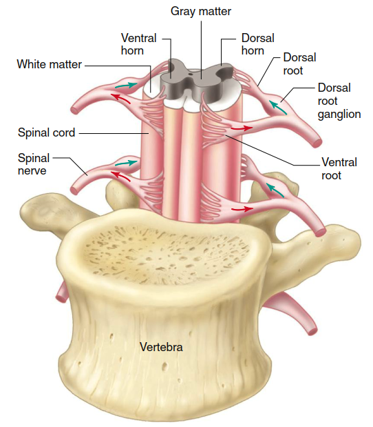
Figure 6.41. Section of the spinal cord, ventral view. The arrows indicate the direction of transmission of neural activity
The gray matter is surrounded by white matter, which comprises groups of myelinated axons. These groups of fiber tracts run longitudinally through the cord: some descending to relay information from the brain to the spinal cord, others ascending to transmit information to the brain. Additional pathways transmit information between different levels of the spinal cord, enabling coordinated segmental responses. This bidirectional conduction system is essential for reflexes, voluntary movement, and sensory perception.
Groups of afferent fibers that enter the spinal cord from the peripheral nerves do so on the dorsal side via the dorsal roots. Small bumps on the dorsal roots, the dorsal root ganglia, contain the cell bodies of these afferent neurons. The axons of efferent neurons leave the spinal cord on the ventral side via the ventral roots. A short distance from the cord, the dorsal and ventral roots from the same level combine to form a spinal nerve, one on each side of the spinal cord, carrying two-way information from both afferent and efferent fibers.
Peripheral Nervous System. Neurons in the peripheral nervous system (PNS) transmit signals between the CNS and receptors and effectors throughout the body. As noted earlier, axons are grouped into bundles called nerves. The PNS contains 43 pairs of nerves: 12 pairs of cranial nerves and 31 pairs of spinal nerves that connect with the spinal cord. Table 6.8 lists the cranial nerves and summarizes the information they transmit. The 31 pairs of spinal nerves are designated by the vertebral levels from which they exit: cervical, thoracic, lumbar, sacral, and coccygeal (see Figure 6.42).
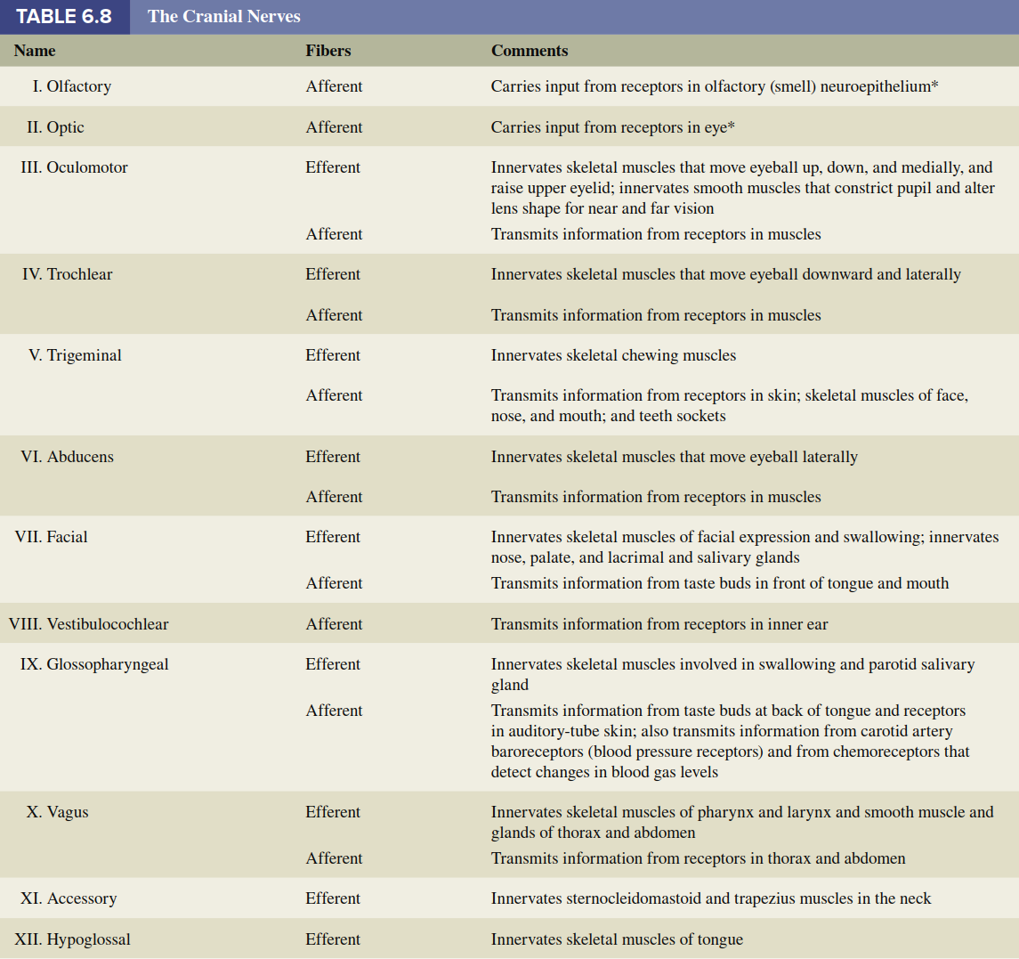
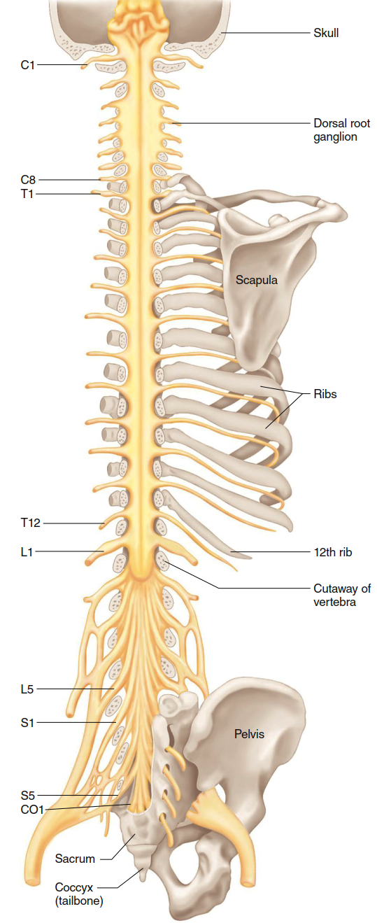
Figure 6.42. Dorsal view of the spinal cord and spinal nerves. Parts of the skull and vertebrae have been cut away; the ventral roots of the spinal nerves are not visible. In general, the eight cervical (C) nerves control the muscles and glands and receive sensory input from the neck, shoulders, arms, and hands. The 12 thoracic (T) nerves are associated with the shoulders, chest, and upper abdomen. The five lumbar nerves (L) are associated with the lower abdomen, hips, and legs; and the five sacral (S) nerves are associated with the genitals and lower digestive tract. The single coccygeal (CO1) nerve innervates the skin region around the tailbone
Neurons in the spinal nerves at each level generally communicate with nearby structures, controlling muscles and glands while receiving sensory input. The eight pairs of cervical nerves innervate the neck, shoulders, arms, and hands. The 12 pairs of thoracic nerves are associated with the chest and upper abdomen. The five pairs of lumbar nerves are associated with the lower abdomen, hips, and legs. The five pairs of sacral nerves are associated with the genitals and lower digestive tract. A single pair of coccygeal nerves associated with the skin over the tailbone region brings the total to 31 pairs.
These peripheral nerves can contain nerve fibers that are axons of efferent neurons, afferent neurons, or both. Therefore, fibers in a nerve may be classified as belonging to the efferent division or the afferent division of the PNS (refer back to Figure 6.37). All spinal nerves contain both afferent and efferent fibers, whereas some cranial nerves contain only afferent fibers (e.g., the optic nerves from the eyes) or only efferent fibers (e.g., the hypoglossal nerve to muscles of the tongue).
As noted earlier, afferent neurons convey information from sensory receptors at their peripheral endings to the CNS. The long portion of their axon lies outside the CNS and is part of the PNS. Afferent neurons are sometimes called primary afferents or first-order neurons because they are the first cells entering the CNS in the synaptically linked chains of neurons that process incoming information. These neurons are pseudounipolar in structure, with their cell bodies residing in dorsal root ganglia or cranial nerve ganglia.
Efferent neurons carry signals from the CNS to muscles, glands, and other tissues. The efferent division of the PNS is more complex than the afferent division, being subdivided into the somatic nervous system and the autonomic nervous system. These terms can be misleading because they suggest additional nervous systems distinct from the central and peripheral systems; however, keep in mind that both subdivisions together constitute the efferent division of the PNS. The autonomic nervous system itself has three branches: sympathetic, parasympathetic, and enteric, each with unique anatomical and functional characteristics.
The simplest distinction between the somatic and autonomic systems is that neurons of the somatic division innervate skeletal muscle, whereas autonomic neurons innervate smooth muscle, cardiac muscle, glands, neurons in the gastrointestinal tract, and other tissues. Additional differences are summarized in Table 6.9, including the number of neurons in the pathway (single for somatic, two-neuron chain for autonomic), the presence of ganglia, and the types of neurotransmitters released.
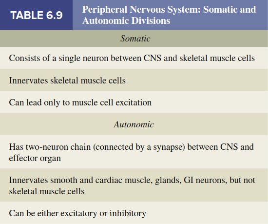
The somatic portion of the efferent division of the PNS comprises all nerve fibers traveling from the CNS to skeletal muscle cells. The cell bodies of these neurons are located in groups within the brainstem or the ventral horn of the spinal cord. Their large-diameter, myelinated axons leave the CNS and pass without any synapses directly to skeletal muscle cells. The neurotransmitter released by these neurons is acetylcholine. Because activity in somatic neurons leads to contraction of the innervated skeletal muscle cells, these neurons are called motor neurons. Excitation of motor neurons produces only contraction of skeletal muscle cells; there are no somatic neurons that directly inhibit skeletal muscles. Instead, muscle relaxation involves the inhibition of motor neurons within the spinal cord via inhibitory interneurons and descending pathways from the brain.
Understanding the structural and functional organization of the spinal cord and peripheral nerves is critical for clinical diagnosis of neurological disorders. Damage to specific spinal roots or peripheral nerves produces characteristic patterns of weakness, sensory loss, or reflex changes. For example, a herniated intervertebral disc may compress a single spinal nerve root, leading to radicular pain and dermatomal sensory deficits. Similarly, peripheral neuropathies often affect distal axons first, producing a "stocking-glove" pattern of sensory loss. These clinical correlations underscore the importance of the anatomical principles described in this section.
Date added: 2026-07-14; views: 140;