
Autonomic Nervous System: Sympathetic and Parasympathetic Divisions
The efferent innervation of non-skeletal muscle tissues occurs through the autonomic nervous system (ANS). A notable exception exists in the gastrointestinal tract, where autonomic neurons synapse onto an intrinsic neuronal network within the organ wall. This complex network, termed the enteric nervous system, contains not only autonomic efferent fibers but also sensory neurons and interneurons. Although frequently categorized as a subdivision of the autonomic efferent system, the enteric nervous system possesses unique integrative capabilities that will be further explored in Chapter 15 within the context of gastrointestinal physiology.
Unlike the somatic nervous system, which uses a single neuron to connect the central nervous system (CNS) to skeletal muscle, the ANS relies on a two-neuron chain linking the CNS with effector cells (Figure 6.43). The first neuron, or preganglionic neuron, has its cell body situated within the CNS, while the synapse between the two neurons occurs outside the CNS in a cluster of cell bodies known as an autonomic ganglion. Neurons projecting from the CNS to the ganglia are designated preganglionic neurons; conversely, those traveling from the ganglia to the target organs are called postganglionic neurons.
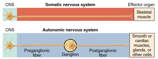
Figure 6.43. Efferent division of the PNS, including an overall plan of the somatic and autonomic nervous systems
Anatomical and physiological distinctions within the ANS form the basis for its further subdivision into the sympathetic and parasympathetic divisions (review Figure 6.37). These two divisions emerge from the CNS at different spinal and cranial levels: sympathetic fibers originate from the thoracic (chest) and lumbar regions of the spinal cord, whereas parasympathetic fibers arise from the brainstem and the sacral portion of the spinal cord (Figure 6.44). Consequently, the sympathetic division is alternatively named the thoracolumbar division, and the parasympathetic division is referred to as the craniosacral division.
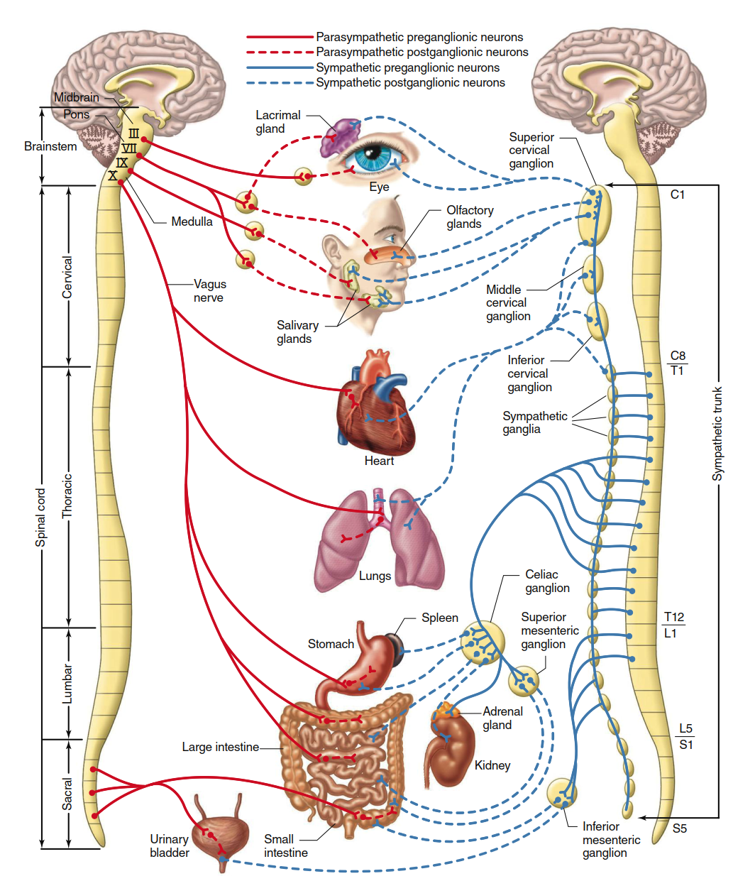
Figure 6.44. The parasympathetic (at left) and sympathetic (at right) divisions of the autonomic nervous system. Although single nerves are shown exiting the brainstem and spinal cord, all represent paired (left and right) nerves. Only one sympathetic trunk is indicated, although there are two, one on each side of the spinal cord. The celiac, superior mesenteric, and inferior mesenteric ganglia are collateral ganglia. Not shown are the fibers passing to the liver, blood vessels, genitalia, and skin glands
The two divisions also display contrasting locations of their autonomic ganglia. Most sympathetic ganglia lie close to the spinal cord and form two parallel chains—one on each side of the vertebral column—known as the sympathetic trunks (see Figure 6.44 and Figure 6.45). Additional sympathetic ganglia, called collateral ganglia (specifically the celiac, superior mesenteric, and inferior mesenteric ganglia), reside within the abdominal cavity, situated nearer to the organs they innervate (see Figure 6.44). In contrast, parasympathetic ganglia are typically located within or immediately adjacent to the target organs that receive postganglionic innervation.
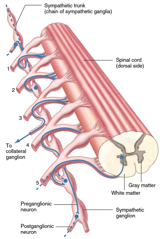
Figure 6.45. Relationship between a sympathetic trunk and spinal nerves (1 through 5) with the various courses that preganglionic sympathetic neurons (solid lines) take through the sympathetic trunk. Dashed lines represent postganglionic neurons. A mirror image of this exists on the opposite side of the spinal cord
Preganglionic sympathetic neurons exit the spinal cord exclusively between the first thoracic and second lumbar segments. Nevertheless, the sympathetic trunks extend the full length of the cord, from cervical levels high in the neck down to sacral levels. Ganglia found in the additional segments of the sympathetic trunks receive preganglionic input from the thoracolumbar region because some preganglionic fibers, upon entering the trunks, travel upward or downward several segments before synapsing with postganglionic neurons (see Figure 6.45, numbers 1 and 4). Other potential pathways taken by sympathetic fibers are illustrated in Figure 6.45, numbers 2, 3, and 5.
The overall activation patterns within the sympathetic versus parasympathetic systems tend to differ significantly. In the sympathetic division, although individual segments may occasionally be activated independently, increased sympathetic activity more commonly occurs body-wide when circumstances demand systemic activation. The parasympathetic system, by contrast, tends to activate specific organs in a highly tailored pattern, precisely matched to each given physiological situation.
In both divisions, acetylcholine (ACh) serves as the neurotransmitter released between preganglionic and postganglionic neurons within autonomic ganglia, and the postganglionic cells predominantly express nicotinic acetylcholine receptors (Figure 6.46). Within the parasympathetic division, ACh is also the neurotransmitter released from the postganglionic neuron onto the effector cell. In the sympathetic division, norepinephrine (NE) usually acts as the transmitter at the postganglionic–effector cell synapse. The qualification “usually” is necessary because a few sympathetic postganglionic endings (e.g., sympathetic pathways regulating sweating) release acetylcholine instead of norepinephrine.
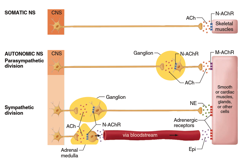
Figure 6.46. Transmitters used in the various components of the peripheral efferent nervous system. Notice that the first neuron exiting the CNS—whether in the somatic or the autonomic nervous system—releases acetylcholine. In a very few cases, postganglionic sympathetic neurons may release a transmitter other than norepinephrine. (ACh, acetylcholine; NE, norepinephrine; Epi, epinephrine; N-AChR, nicotinic acetylcholine receptor; M-AChR, muscarinic acetylcholine receptor)
Beyond the classical autonomic neurotransmitters described above, a widespread network of postganglionic neurons has been identified as nonadrenergic and noncholinergic (NANC). These neurons release nitric oxide (NO) and other signaling molecules to mediate certain forms of blood vessel dilation and to regulate various gastrointestinal, respiratory, urinary, and reproductive functions.
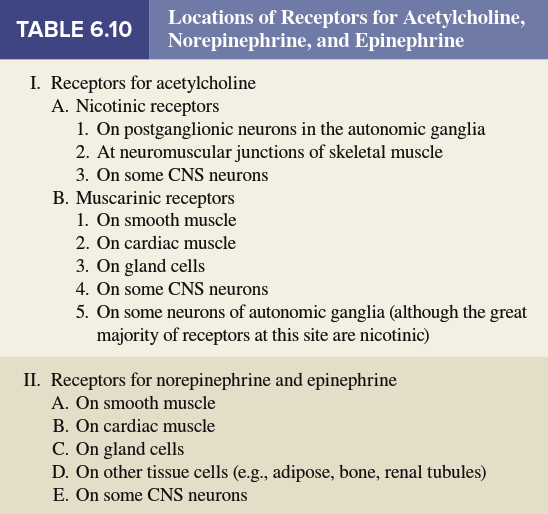
Numerous drugs that stimulate or inhibit components of the ANS exert their effects by targeting receptors for acetylcholine and norepinephrine. It is important to recall that multiple receptor subtypes exist for each neurotransmitter. The vast majority of acetylcholine receptors in autonomic ganglia are nicotinic receptors. In contrast, the acetylcholine receptors found on cellular targets of postganglionic autonomic neurons are muscarinic receptors (Table 6.10). (Note that cholinergic receptors on skeletal muscle fibers—innervated by somatic motor neurons, not autonomic neurons—are also nicotinic receptors, but they differ in subunit composition and pharmacology.)
One unique subset of postganglionic neurons in the sympathetic division never develops axons. Instead, these neurons form part of an endocrine gland known as the adrenal medulla (see Figure 6.46). When activated by preganglionic sympathetic axons, adrenal medullary cells release a mixture of approximately 80% epinephrine and 20% norepinephrine directly into the bloodstream. Under these circumstances, these catecholamines are properly termed hormones rather than neurotransmitters because they are transported via the blood to effector cells that possess appropriate receptors. Some of these receptors are identical to the adrenergic receptors located near sympathetic postganglionic release sites and are normally activated by neuron-derived norepinephrine. However, other receptors reside in locations distant from sympathetic nerve endings and are therefore activated only by circulating epinephrine or norepinephrine. The overall physiological effects of the two catecholamines differ slightly because certain adrenergic receptor subtypes (e.g., β2 receptors) have a higher affinity for epinephrine, whereas others (e.g., α1 receptors) show greater affinity for norepinephrine.
Table 6.11 provides a reference summary of the effects of autonomic nervous system activity, which will be elaborated in later chapters. Notably, the heart, many glands, and various smooth muscles receive dual innervation from both sympathetic and parasympathetic fibers. In most cases, the effect elicited by one division on a given effector cell is opposite to that produced by the other division (several exceptions to this rule are indicated in Table 6.11). Moreover, the two divisions are typically activated reciprocally: as the activity of one division increases, the activity of the other decreases. This reciprocal relationship can be likened to driving a car with one foot on the brake and the other on the accelerator. Either depressing the brake (parasympathetic activation) or releasing the accelerator (sympathetic withdrawal) will slow the vehicle. Dual innervation by antagonistic neurons provides exquisitely fine control over effector organs, representing a prime example of the general physiological principle that most functions are regulated by multiple, often opposing, systems.
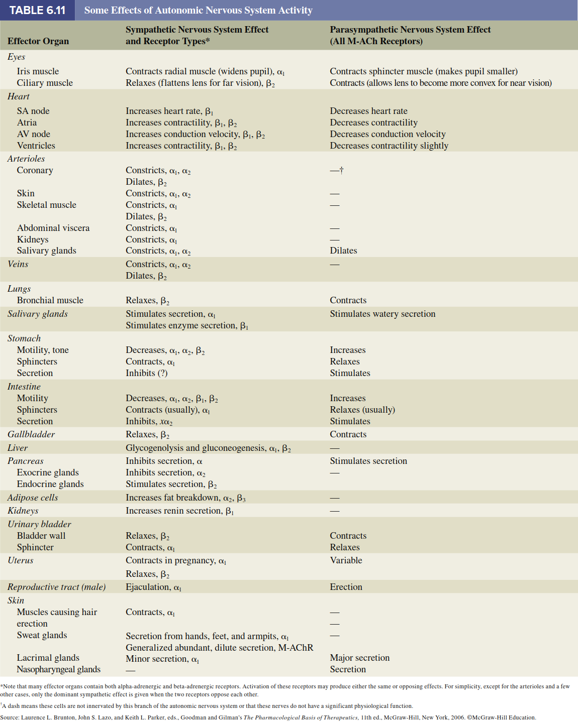
A useful generalization is that the sympathetic system increases its activity during periods of physical or psychological stress. Indeed, a generalized, body-wide activation of the sympathetic division is known as the fight-or-flight response, which describes the situation of an animal preparing either to confront an attacker or to flee from it. All resources supporting physical exertion are mobilized: heart rate and blood pressure rise, blood flow increases to skeletal muscles, the heart, and the brain, the liver releases glucose, and the pupils dilate. Simultaneously, sympathetic firing inhibits gastrointestinal activity and reduces blood flow to the digestive tract. In contrast, when the parasympathetic system is activated, a person enters a rest-or-digest state, in which most of the above processes are reversed or remain inactive.
The two divisions of the autonomic nervous system rarely operate in isolation, and most autonomic responses represent the regulated interplay of both sympathetic and parasympathetic inputs. This integrated control allows the body to respond dynamically to ever-changing internal and external environments, maintaining homeostasis through balanced autonomic outflow.
Date added: 2026-07-14; views: 156;