
Central and Peripheral Nervous System: Anatomy, Functions, and Brain Structures Explained
The anatomy and broad functions of the major structures within the central nervous system (CNS) and peripheral nervous system (PNS) are surveyed in this section. Figure 6.37 provides a conceptual overview of the organization of the nervous system, which serves as a reference for discussing various subdivisions in this section and later chapters. Understanding these structural relationships is fundamental to comprehending neural signal processing and integration.
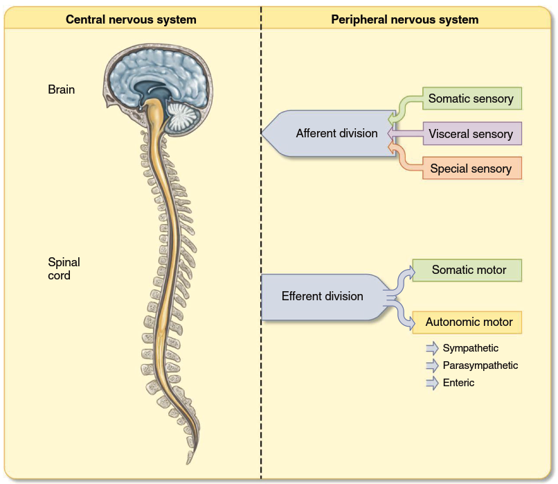
Figure 6.37. Overview of the structural and functional organization of the nervous system
Key terminology must first be introduced to facilitate accurate communication. A long extension from a single neuron is called an axon or nerve fiber, whereas the term nerve refers to a group of many axons traveling together to and from the same general location within the PNS. Notably, there are no nerves in the CNS; instead, a group of axons traveling together in the CNS is called a pathway, a tract, or, when connecting the right and left halves of the brain, a commissure. Two general types of pathways occur in the CNS: long neural pathways consist of neurons with relatively long axons that carry information directly between the brain and spinal cord or between large brain regions, whereas multisynaptic pathways include many neurons with branching axons and numerous synaptic connections. Because synapses are sites where new information can be integrated into neural messages, multisynaptic pathways perform complex neural processing, while long neural pathways transmit signals with relatively less alteration.
Clusters of neuron cell bodies with similar functions are commonly observed throughout the nervous system. In the PNS, these clusters are called ganglia (singular, ganglion), while in the CNS they are termed nuclei (singular, nucleus) — a term not to be confused with the cell nucleus. This organizational principle allows for efficient processing and relay of neural information across different anatomical regions.
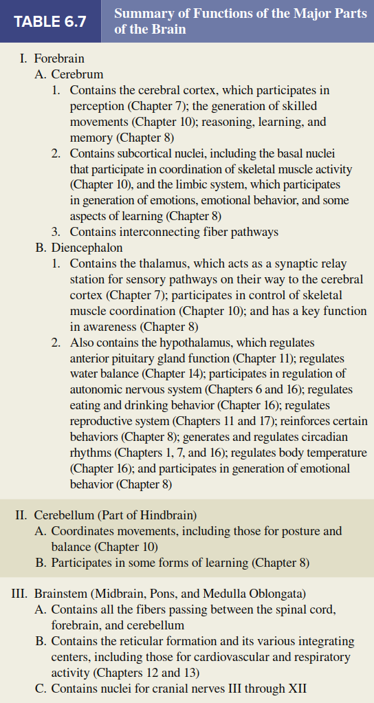
Central Nervous System: Brain. During development, the CNS forms from a long tube. As the anterior part of this tube — which becomes the brain — folds during its continuing formation, three distinct regions initially become apparent: the forebrain, midbrain, and hindbrain (see Figure 6.38). These regions continue to develop, forming further subdivisions: the forebrain develops into the cerebrum and diencephalon; the midbrain remains as a single major division; and the hindbrain develops into the pons, medulla oblongata, and cerebellum. The pons, medulla oblongata, and midbrain are heavily interconnected and share many similar functions; due to this and their anatomical location, they are considered together as the brainstem. The brain also contains four interconnected cavities, the cerebral ventricles, which are filled with fluid and provide physical support for the brain. Overviews of the brain subdivisions are included here and in Table 6.7, though detailed functions are more fully presented in Chapters 7, 8, and 10.
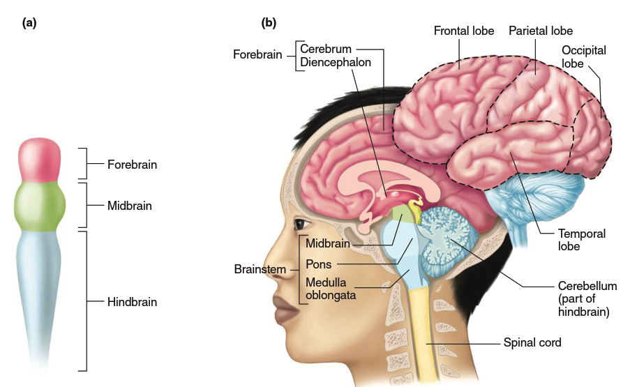
Figure 6.38. Structures of the human brain. (a) Development of the three major parts of the brain in a 4-week-old embryo. (b) The major divisions of the adult brain shown in sagittal section. The outer surface of the cerebrum (cortex) is divided into four lobes as shown
Forebrain: The Cerebrum. The larger component of the forebrain, the cerebrum, consists of the right and left cerebral hemispheres as well as associated structures on the underside of the brain. The cerebral hemispheres (Figure 6.39) consist of the cerebral cortex — an outer shell of gray matter composed primarily of cell bodies that give the area a gray appearance — and an inner layer of white matter composed primarily of myelinated fiber tracts. The cerebral cortex overlies cell clusters, also gray matter, collectively termed the subcortical nuclei. The fiber tracts consist of many nerve fibers that bring information into the cerebrum, carry information out, and connect different areas within a hemisphere. Although largely separated by a deep longitudinal division, the cortex layers of the left and right cerebral hemispheres are connected by a massive bundle of nerve fibers known as the corpus callosum.
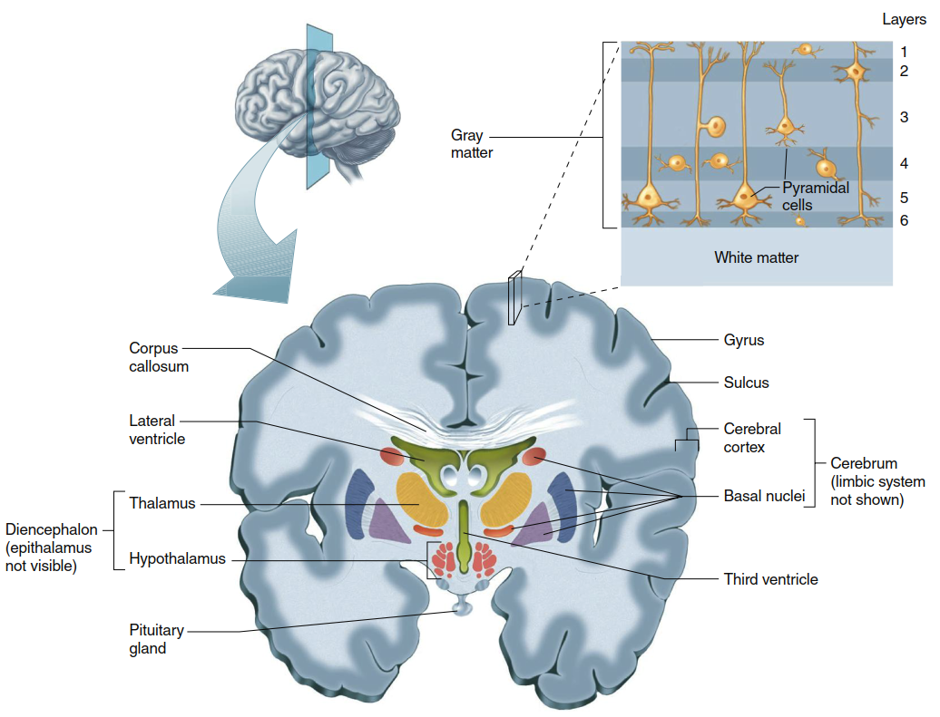
Figure 6.39. Frontal section of the cerebral hemispheres showing portions of the cerebrum and underlying diencephalon (thalamus and hypothalamus; the epithalamus is not visible in this plane of section). The limbic system is shown in Figure 6.40. The corpus callosum is a fiber tract that connects the two hemispheres, which are folded into gyri and sulci. Some of the fluid-filled ventricles of the brain are also indicated, as is the pituitary gland. The inset shows a simplified depiction of the six-layer organization of the cerebral cortex. Not shown is the extensive degree of neuronal input into the different layers from cells outside the cerebral cortex
Cerebral Cortex. The cerebral cortex of each cerebral hemisphere is divided into four lobes, named after the overlying skull bones: frontal, parietal, occipital, and temporal lobes. Although averaging only 3 mm in thickness, the cerebral cortex is highly folded, resulting in an area containing cortical neurons that is four times larger than it would be without folding, yet without appreciably increasing brain volume. This folding produces the characteristic external appearance of the human cerebrum, with sinuous ridges called gyri (singular, gyrus) separated by grooves called sulci (singular, sulcus).
The cells of the human cerebral cortex are organized in six distinct layers, composed of varying sizes and numbers of two basic cell types: pyramidal cells (named for the shape of their cell bodies) and nonpyramidal cells. Pyramidal cells form the major output cells of the cerebral cortex, sending their axons to other parts of the cortex and to other CNS regions. Nonpyramidal cells are mostly involved in receiving inputs into the cerebral cortex and in local information processing. This elaboration into multiple cell layers, like the highly folded structure, allows for increased number and integration of neurons for signal processing. Such specialization of structural surface area to enhance function affirms the general principle of physiology that structure and function are related. Indeed, an increase in the number of cell layers in the cerebral cortex has paralleled the increase in behavioral and cognitive complexity in vertebrate evolution — for example, reptiles have just three layers, dolphins have five, and some regions of the human brain with ancient evolutionary origins (such as the olfactory cortex) persist in having only three cell layers.
The cerebral cortex is one of the most complex integrating areas of the nervous system. Here, basic afferent information is collected and processed into meaningful perceptual images, and control over systems that govern skeletal muscle movement is refined. Nerve fibers enter the cerebral cortex predominantly from the diencephalon and brainstem areas; extensive signaling also occurs between areas within the cerebral cortex. Some input fibers convey information about specific environmental events, whereas others control levels of cortical excitability, determine states of arousal, and direct attention to specific stimuli.
Basal Nuclei. The subcortical nuclei are heterogeneous groups of gray matter lying deep within the cerebral hemispheres. Predominant among them are the basal nuclei (often, though less correctly, referred to as basal ganglia), which have important functions in controlling movement and posture as well as more complex aspects of behavior.
Limbic System. Thus far, discrete anatomical areas of the forebrain have been described. Some of these forebrain areas, consisting of both gray and white matter, are also classified together in a functional system called the limbic system. This interconnected group of brain structures includes portions of frontal-lobe cortex, temporal lobe, thalamus, and hypothalamus, as well as the fiber pathways that connect them (Figure 6.40). Besides being connected with each other, the parts of the limbic system connect with many other CNS regions. Structures within the limbic system are associated with learning, emotional experience and behavior, and a wide variety of visceral and endocrine functions (see Chapter 8).
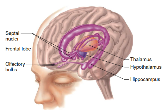
Figure 6.40. Major structures of the limbic system (portions enhanced in violet) and their anatomical relation to the hypothalamus (purple) are shown in this partially transparent view of the brain
Forebrain: The Diencephalon. The diencephalon, divided in two by the narrow third cerebral ventricle, is the second component of the forebrain. It contains the thalamus, hypothalamus, and epithalamus (see Figure 6.39). The thalamus is a collection of several large nuclei that serve as synaptic relay stations and important integrating centers for most inputs to the cortex, and it has a key function in general arousal. The thalamus is also involved in focusing attention; for example, it is responsible for filtering out extraneous sensory information, such as might occur when trying to concentrate on a private conversation at a loud, crowded party.
The hypothalamus lies below the thalamus on the undersurface of the brain and, like the thalamus, contains numerous different nuclei. These nuclei and their pathways form the master command center for neural and endocrine coordination. Indeed, the hypothalamus is the single most important control area for homeostatic regulation of the internal environment. Behaviors related to preservation of the individual (e.g., eating and drinking) and preservation of the species (reproduction) are among the many functions of the hypothalamus. The hypothalamus lies directly above and is connected by a stalk to the pituitary gland, an important endocrine structure that the hypothalamus regulates (Chapter 11). As mentioned earlier, some parts of the hypothalamus and thalamus are also considered part of the limbic system.
The epithalamus is a small mass of tissue that includes the pineal gland, which participates in the control of circadian rhythms through release of the hormone melatonin.
Hindbrain: The Cerebellum. The cerebellum consists of an outer layer of cells, the cerebellar cortex (not to be confused with the cerebral cortex), and several deeper cell clusters. Although the cerebellum does not initiate voluntary movements, it is an important center for coordinating movements and for controlling posture and balance. To carry out these functions, the cerebellum receives information from muscles, joints, skin, eyes, vestibular apparatus, viscera, and the parts of the brain involved in movement control. Although the cerebellum’s function is almost exclusively motor, recent research strongly suggests that it may also be involved in some forms of learning. The other hindbrain components — the pons and medulla oblongata — are considered together with the midbrain as part of the brainstem.
Brainstem: The Midbrain, Pons, and Medulla Oblongata. All nerve fibers that relay signals between the forebrain, cerebellum, and spinal cord pass through the brainstem. Running through the core of the brainstem and consisting of loosely arranged nuclei intermingled with bundles of axons is the reticular formation — the one part of the brain absolutely essential for life. It receives and integrates input from all CNS regions and processes a great deal of neural information. The reticular formation is involved in motor functions, cardiovascular and respiratory control, and the mechanisms that regulate sleep, wakefulness, and attention. Most of the biogenic amine neurotransmitters are released from axons of cells in the reticular formation; due to the far-reaching projections of these cells, these neurotransmitters affect all levels of the nervous system.
Pathways that convey information from the reticular formation to upper brain regions stimulate arousal and wakefulness. They also direct attention to specific events by selectively stimulating neurons in some brain areas while inhibiting others. Fibers that descend from the reticular formation to the spinal cord influence activity in both efferent and afferent neurons. Considerable interaction occurs between reticular pathways that go up to the forebrain, down to the spinal cord, and to the cerebellum; for example, all three components function together in controlling muscle activity.
The reticular formation encompasses a large portion of the brainstem, and many areas within it serve distinct functions. For instance, some reticular formation neurons are clustered together, forming brainstem nuclei and integrating centers, including the cardiovascular, respiratory, swallowing, and vomiting centers (discussed in later chapters). The reticular formation also has nuclei important in eye-movement control and the reflexive orientation of the body in space. In addition, the brainstem contains nuclei involved in processing information for 10 of the 12 pairs of cranial nerves — the peripheral nerves that connect directly with the brain and innervate the muscles, glands, and sensory receptors of the head, as well as many organs in the thoracic and abdominal cavities.
Date added: 2026-07-14; views: 141;