The human chromosome diseases
Using of the cytogenetic method makes it possible to separate groups of diseases related with imbalance in chromosome number and structure as well. They are called Human chromosome diseases. Statistically, it was determined that 0.7% of newborns have chromosomal diseases. Deviance in chromosome number is related with chromosomes separation in meiosis. The deviance in sex chromosomes is not lethal, but they often lead to decreased fertility and some development abnormalities.
There are the following human sex chromosome diseases.
Additional Y-chromosome has less severe effects on phenotype. There is no special sign to distinguish person having additional Y-chromosome. It is known that part of them develop pattern of antisocial development. The most of men having additional Y-chromosome are fertile. It makes genetic analysis of them more complicated.
Additional X-chromosome in women. It gives a wide phenotypical polymorphism. It occurs with rate 1.4:1000 girls. Diagnostic feature is having two sex chromatin bodies in buccal epithelium cells. The most of individuals having genotype 47, XXX express normal physical and mental phenotype without any deviations in reproductive system. But some of them may have pathological changes in reproductive system such as secondary anemia, dismenorrhea, early climax. Intellectual development is normal or on a lowest limit of normal condition. It was founded that there is a higher rate of schizophrenia among X-chromosome tri-somic women. In rare cases, such as X-chromosome polysomy, the deviations are more expressed.
Additional X-chromosome in men (Kleinefelter ’s syndrome). It gives a wide phenotypical polymorphism. It occurs in about 1 out 500 male births. The typical feature is a having of sex chromatin in nucleus of buccal epithelial cells. It becomes evident in puberty. The clinical signs mainly concerns insufficient development of male secondary sexual characteristics. They are tall, with long limbs, with sclerotic degeneration of semeniferous tubules and, in some cases with diminished mental capacity. The XXY complex does not lead to perinatal death. However, The XXY complex is founded in perinatal kid’s deaths 10 times more often than in survived children (pic 11.3).
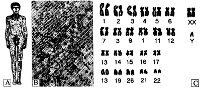
Pic. 11.3. The Kleinefelter’s syndrome (A) with typical testis histology (B) and idiogramm (C) (by M. L. Barr, 1948, E. Bergemann, 1962)
Absence of X-chromosome in women (Turner 's syndrome). It occurs roughly once in every 5000 female births. Such individuals have no sex chromatin in nucleus of buccal epithelial cells. It results in a sterile female of short stature, a webbed neck, disk shaped thorax, and immature sex organs that do not undergo puberty changes. Sometimes they have defects of color perception. Such embryos are subjected to high prenatal mortality, that why their population rate is small (pic 11.4).

Pic. 11.4. The Turner’s syndrome: The girl with syndrome and her chromosome set (by O. belong et al., 1963)
Absence of X-chromosome in men. Such zygote is enviable and fails to develop further. The humans can not survive without any of the genes on the X- chromosome.
Sex chromosome aberrations. The most common is deletion of short arm of X-chromosome. It leads to formation of phenotype similar as X-chromosome monosomy.
Among autosomal set changes there are following which are most common.
Trisomy 21 (Down syndrome). The individuals having the syndrome have decreased size of scalp, small in stature, poor muscle tone, big gap between I and II finger of foot, immatured sex organs, mental retardation (pic 11.5). The mental retardation pathogenesis of trisomy 21 includes central nervous system immaturation, in particular, the insufficient myelinization of nervous fibers. About half have heart defects and defects of big vessels. The Down syndrome rate is about one out of700-800 births. The average age of mothers having children with Down syndrome is on average 6-8 years older then the age of mothers having normal children. The life span of such individuals is about 21-24 years.
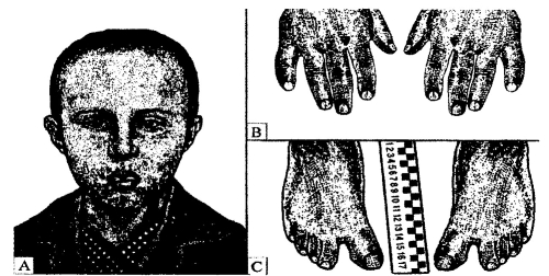
Pic. 11.5. The Down syndrome: A - patient of 13 years old; В - small finger deviation; C - big gap between I and II finger of foot (by E. F. Davidenkova, 1965)
Trisomy 18 (Edward’s syndrome). It is third in rate after trisomy 21 and 13. Individuals have severe prenatal immaturation and numerous defects of skeletal system, in particular, facial part of scalp. The internal defects are defects of interventriculum septum of heart, defects of aortic valve and pulmonary artery valve, cryptorchysm in males. They also have severe mental retardation, abnormal bending of joints, and prevalating length of index finger over middle one, low ear’s position, and small lower jaw. With a good treatment they can survive till one year of age (pic 10.6).
Trisomy 13 (Pattaw ’s syndrome). Its rate is 1:5000 -1:7000. The trisomic 13 individuals die in early childhood. More than 90% die in first year of life. Individuals have defects of brain and scalp (pic 10.7). The second group of defects is defects in finger number - polydactilia, especially hexodactilia (having 6 fingers). They also have , abnormal bending of joints, defects of heart septa, incomplete intestine turn, abnormalities in inner reproductive organs in both sex children, typical changes in pancreas (by Lasuc G.1.1979). Some embryos with trisomy 13 are subjected to high prenatal mortality, that why their population rate is small.
Autosome aberrations. The most common are deletions of 5th and 18th chromosomes. Deletion of 5th chromosome short arm was described by J. Lejen as “cat”s scream” syndrome. The child’s scream songs like a cat’s scream. Other symptoms are larynx immaturation, microcefalia, mental retardation, poor muscle tone, low ear’s position, and underdeveloped sexual characteristics. Deletion of long or short arm of 18 chromosome leads to face defects, skeletal defects, internal defects, microcephalia, mental retardation and other abnormalities.
Different translocations. The provide development of different chromosome diseases. They can be of such variants: translocation of 21 chromosomes to 15 chromosomes resulting in Down syndrome, translocation of 21 chromosomes to 13,14 and 22 chromosome.
The rate of chromosome abnormalities corresponds with mother age, starting from 35.
Date added: 2023-01-09; views: 758;