
Glial Cells, Neural Growth, and Regeneration in the Human CNS
According to recent analyses, neurons constitute only about half of the cells in the human central nervous system (CNS). The remainder are glial cells (glia, meaning “glue”), which surround the axons and dendrites of neurons and provide physical and metabolic support. Unlike most neurons, glial cells retain the capacity to divide throughout life. Consequently, many CNS tumors originate from glial cells rather than from neurons.
Several types of glial cells are found in the CNS (Figure 6.6). One type discussed earlier is the oligodendrocyte, which forms the myelin sheath of CNS axons. A second type, the astrocyte, helps regulate the composition of extracellular fluid in the CNS by removing potassium ions and neurotransmitters around synapses. Astrocytes also stimulate the formation of tight junctions (review Figure 3.9) between capillary wall cells in the CNS, creating the blood‑brain barrier—a much more selective filter for exchanged substances than exists between blood and most other tissues.
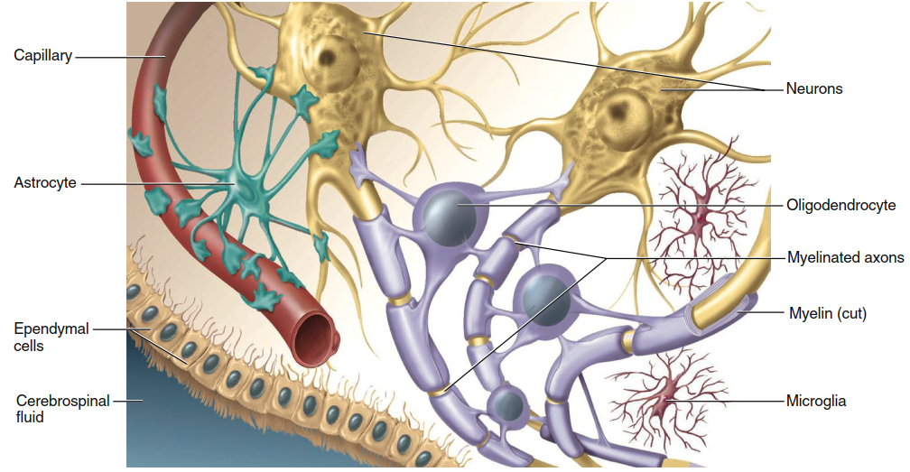
Figure 6.6. Glial cells of the central nervous system
Astrocytes sustain neurons metabolically by providing glucose and removing the waste product ammonia. In embryos, they guide migrating CNS neurons to their final destinations and stimulate neuronal growth by secreting growth factors. Additionally, astrocytes possess many neuron‑like characteristics, including ion channels, receptors for certain neurotransmitters with their processing enzymes, and the ability to generate weak electrical responses. Thus, beyond their well‑defined functions, astrocytes are speculated to participate in information signaling within the brain.
The microglia, a third type of CNS glial cell, are specialized macrophage‑like cells that perform immune functions in the CNS and may also contribute to synapse remodeling and plasticity. Lastly, ependymal cells line the fluid‑filled cavities within the brain and spinal cord, regulating the production and flow of cerebrospinal fluid (described later). Schwann cells, the glial cells of the peripheral nervous system (PNS), share most properties of CNS glia. As mentioned earlier, Schwann cells produce the myelin sheath of peripheral neuron axons.
Neural Growth and Development. The elaborate networks of neuronal processes depend upon the outgrowth of specific axons to specific targets. Development of the nervous system in the embryo begins with divisions of undifferentiated precursor cells (stem cells) that can develop into neurons or glia. After the final cell division, each neuronal daughter cell differentiates, migrates to its final location, and extends processes that will become its axon and dendrites. A specialized enlargement called the growth cone forms the tip of each extending axon and is involved in finding the correct route and final target.
As the axon grows, it is guided along the surfaces of other cells, most commonly glial cells. The route followed depends largely on attracting, supporting, deflecting, or inhibiting influences exerted by several types of molecules. Some of these molecules, such as cell adhesion molecules, reside on the membranes of glia and embryonic neurons. Others are soluble neurotrophic factors (growth factors for neural tissue) in the extracellular fluid surrounding the growth cone or its distant target.
Once the target of the advancing growth cone is reached, synapses form. During these early stages of neural development—occurring throughout all trimesters of pregnancy and into infancy—alcohol, other drugs, radiation, malnutrition, and viruses can cause permanent damage to the developing fetal nervous system. A surprising aspect of neural development occurs after growth and projection of the axons: many newly formed neurons and synapses degenerate. In fact, 50% to 70% of neurons undergo programmed self‑destruction called apoptosis in the developing CNS. Although the reason for this seemingly wasteful process is unknown, neuroscientists speculate that it refines or fine‑tunes connectivity.
Throughout life, the brain exhibits an remarkable ability to modify its structure and function in response to stimulation or injury, a characteristic known as plasticity. This may involve the generation of new neurons but particularly involves the remodeling of synaptic connections. These events are stimulated by exercise and by engaging in cognitively challenging activities. The degree of neural plasticity varies with age.
For many neural systems, a critical time window for development occurs at a young age. In visual pathways, for example, brain regions involved in processing visual stimuli are permanently impaired if no visual stimulation is received during a critical period that peaks between 1 and 2 years of age. By contrast, the ability to learn a language undergoes a slower, more subtle change in plasticity—humans learn languages relatively easily until adolescence, but learning becomes slower and more difficult from adolescence through adulthood.
The basic shapes and locations of major neuronal circuits in the mature CNS do not change once formed. However, the creation and removal of synaptic contacts begun during fetal development continue throughout life as part of normal growth, learning, and aging. Although it was previously thought that new neuron production ceased around birth, a growing body of evidence indicates that the ability to produce new neurons is retained in some brain regions throughout life. For example, cognitive stimulation and exercise increase neuron numbers in brain regions associated with learning, even in adults. Additionally, the effectiveness of some antidepressant medications depends upon the production of new neurons in regions involved in emotion and motivation.
Regeneration of Axons. If axons are severed, they can repair themselves and restore significant function provided the damage occurs outside the CNS and does not affect the neuron’s cell body. After such an injury, the axon segment separated from the cell body degenerates. The part of the axon still attached to the cell body then gives rise to a growth cone, which grows out to the effector organ so that function can be restored. Return of function following a peripheral nerve injury is delayed because axon regrowth proceeds at only about 1 mm per day. For instance, if afferent neurons from the thumb were damaged near the shoulder, it might take two years for thumb sensation to be restored.
Spinal injuries typically crush rather than cut the tissue, leaving axons intact. In this case, a primary problem is apoptosis of nearby oligodendrocytes. When these cells die and their associated axons lose their myelin sheath, the axons cannot transmit information effectively. Severed axons within the CNS may grow small new extensions, but no significant regeneration occurs across the damaged site, and there are no well‑documented reports of substantial return of function. Functional regeneration is prevented either by some basic difference of CNS neurons or by properties of their environment, such as inhibitory factors from nearby glia. Presumably, evolutionary selection pressure limited growth of neurons in the mature CNS to minimize disruption of the precise architecture of complex neuronal networks.
Researchers are testing various ways to support axonal regeneration in the CNS. They are creating tubes to support regrowth of severed axons, redirecting axons to spinal cord regions lacking growth‑inhibiting factors, preventing oligodendrocyte apoptosis to maintain myelin, and supplying neurotrophic factors that support tissue recovery. Medical researchers are also attempting to restore function to damaged spinal cords and brains by implanting undifferentiated stem cells that develop into new neurons and replace missing neurotransmitters or neurotrophic factors. Initial stem cell research focused on embryonic and fetal stem cells, which, while promising, raise ethical concerns. Recently, however, researchers have developed promising techniques using stem cells isolated from adults, as well as adult cells induced to revert to a stem‑cell‑like state.
Date added: 2026-07-14; views: 143;